Japanese
English
- 有料閲覧
- Abstract 文献概要
- 1ページ目 Look Inside
I.はじめに
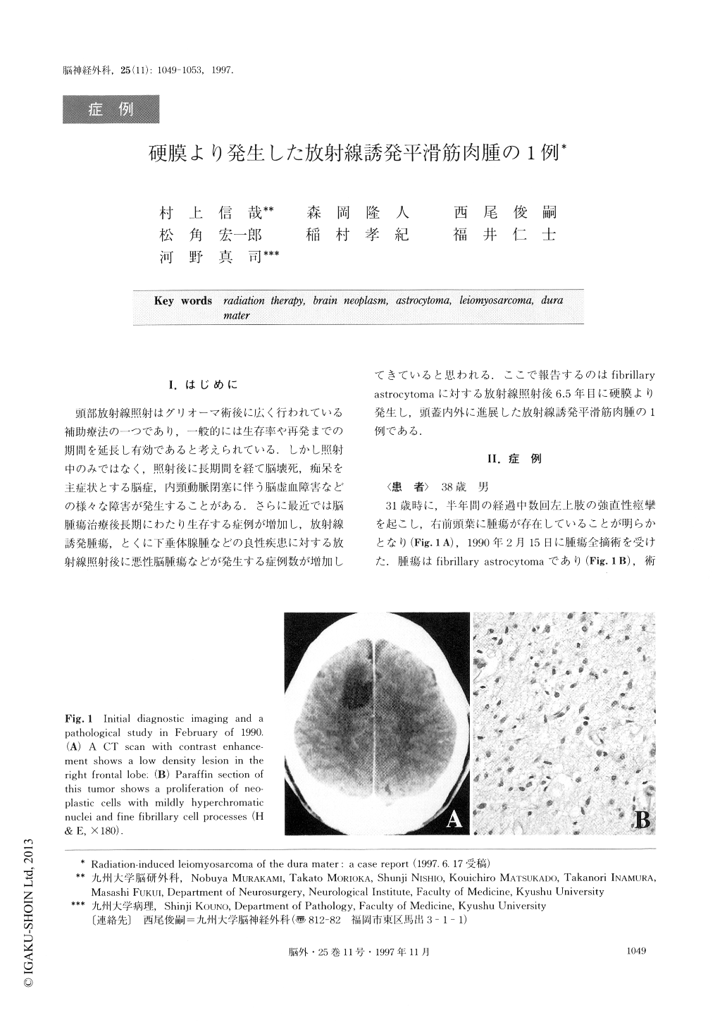
頭部放射線照射はグリオーマ術後に広く行われている補助療法の一つであり,一般的には生存率や再発までの期間を延長し有効であると考えられている.しかし照射中のみではなく,照射後に長期間を経て脳壊死,痴呆を主症状とする脳症,内頸動脈閉塞に伴う脳虚血障害などの様々な障害が発生することがある.さらに最近では脳腫瘍治療後長期にわたり生存する症例が増加し,放射線誘発腫瘍,とくに下垂体腺腫などの良性疾患に対する放射線照射後に悪性脳腫瘍などが発生する症例数が増加してきていると思われる.ここで報告するのはfibrillary astrocytomaに対する放射線照射後6.5年目に硬膜より発生し,頭蓋内外に進展した放射線誘発平滑筋肉腫の1例である.
A 31-year-old man underwent total resection for a fibrillary astrocytoma in the right frontal lobe followed by 6MV x-ray radiotherapy. The portal field size was a square of 8cm×7cm, and the total dose of irradiation was 50Gy, with single fractions of 2Gy. For the next 6.5 years there was no recurrence of the astrocytoma. At 38 years of age, the patient noticed a subcutaneous mass in the scar of the previous operation and de-veloped generalized convulsive seizures. MRI revealed a dural tumor within the previous radiation field, and the tumor was partially removed. Histologically, it was diagnosed as a leiomyosarcoma. This dural sarcoma satisfies the widely used criteria for definition of radia-tion-induced malignancies first described by Cahan et al. Both the clinical features and the possible histogene-sis of this secondary tumor are briefly discussed.

Copyright © 1997, Igaku-Shoin Ltd. All rights reserved.


