Japanese
English
- 有料閲覧
- Abstract 文献概要
- 1ページ目 Look Inside
I.はじめに
FLAIR法(fluid attenuated inversion recovery sequ—ence)は,自由水の信号を抑制したT2強調画像が得られるMRIの撮像法の一種である1-17).脳梗塞とWir—chow Robin腔との鑑別2,4,12-14)や,脊髄病変の描出6,15,17)などでの有用性が指摘されているが,脳腫瘍像に関してはいまだ報告は少ない4,12).
われわれは,高速FLAIR法を種々の頭蓋内腫瘍34症例に適応し,詳細に読影した上で従来の撮像法の画像と比較した.その結果FLAIR法は腫瘍摘出術を施行する上で極めて有用な撮像法であると思われたので以下に詳述する.
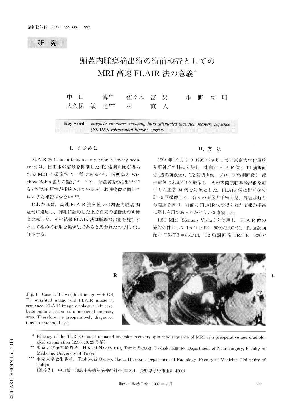
Whenever the extirpation of intracranial tumors is planned, neurosurgeons always keep their eyes on the cerebrospinal fluid (CSF) space around intracranial tumors. If enough space exists in the neighborhood of the tumors, the damage to adjacent parenchyma may be reduced by the procedure through the CSF space. A newly advanced MRI pulse sequence: the FLAIR (fluid attenuated inversion recovery) imaging, in which a long TE spin echo sequence is used with suppression of the CSF with an inversion pulse, displays the CSF space as a no-signal intensity area. There have been only a few reports, however, on the FLAIR pulse sequ-ence of brain tumors as yet. We examined 34 cases of intracranial tumors by FLAIR images and analyzed the advantages and disadvantages of the FLAIR pulsesequence for decision making on tumor removal.
Making use of the FLAIR pulse sequence, the CSF space is depicted as a no-signal intensity area and much more information about perifocal edema and the inva-sion area around the tumors can be provided than that provided by the other ordinary pulse sequences (T1 weighted images, T2 weighted images and Proton weighted images). Therefore, operative strategies can be more easily worked out on the FLAIR images. Furthermore, the difference between arachnoid and epidermoid is able to be detected on the FLAIR images.
Nevertheless, on FLAIR images, the tumors withoutperifocal edema or invasion to adjacent parenchyma were not apparent and the difference between tumoral dissemination into multi-ventricular space and the periventriculuar artifact of FLAIR images could not be distinguished. The FLAIR pulse sequence has other artifacts like intraventricular flow related enhancement and so on.
If the images are carefully checked up on the above-mentioned points, the FLAIR pulse sequence of MRI can not fail to be useful in making plans for operations on intracranial neoplasms.

Copyright © 1997, Igaku-Shoin Ltd. All rights reserved.


