Japanese
English
- 有料閲覧
- Abstract 文献概要
- 1ページ目 Look Inside
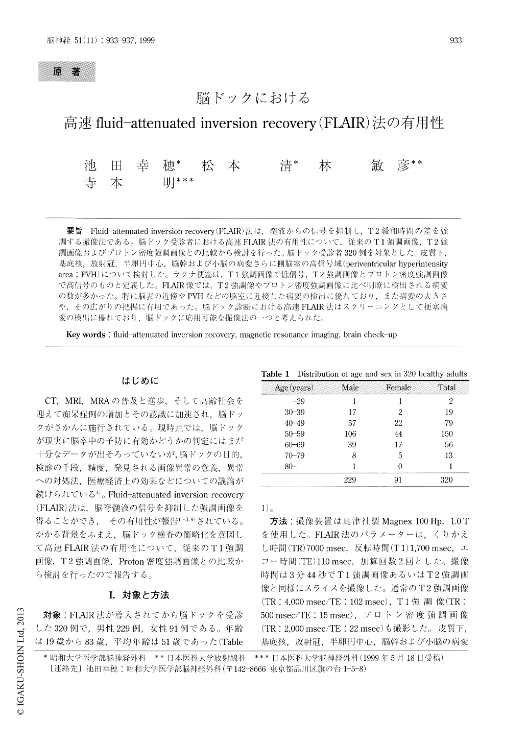
Fluid-attenuated inversion recovery(FLAIR)法は,髄液からの信号を抑制し,T2緩和時間の差を強調する撮像法である。脳ドック受診者における高速FLAIR法の有用性について,従来のT1強調画像,T2強調画像およびプロトン密度強調画像との比較から検討を行った。脳ドック受診者320例を対象とした。皮質下,基底核,放射冠,半卵円中心,脳幹および小脳の病変さらに側脳室の高信号域(periventricular hyperintensityarea;PVH)について検討した。ラクナ梗塞は,T1強調画像で低信号,T2強調画像とプロトン密度強調画像で高信号のものと定義した。FLAIR像では,T2強調像やプロトン密度強調画像に比べ明瞭に検出される病変の数が多かった。特に脳表の近傍やPVHなどの脳室に近接した病変の検出に優れており,また病変の大きさや,その広がりの把握に有用であった。脳ドック診断における高速FLAIR法はスクリーニングとして梗塞病変の検出に優れており,脳ドックに応用可能な撮像法の一つと考えられた。
Fluid-attenuated inversion recovery (FLAIR) images are magnetic resonance (MR) images obtained with an inversion recovery sequence having a long inversion time (T1) and a long echo time (TE). The purpose of this study was to evaluate the utility of FLAIR images in brain check-up. 320 healthy adults (229 males, 91 females) were examined with FLAIR sequences of several types having repetitive time (TR) of 7000 msec, inversion times of 1700 msec and echo times (TE) of 110 msec. All studies were performed on a SHIMAZU MAGNEX 100 HP 1.0 T imaging system.

Copyright © 1999, Igaku-Shoin Ltd. All rights reserved.


