Japanese
English
- 有料閲覧
- Abstract 文献概要
- 1ページ目 Look Inside
- 参考文献 Reference
Ⅰ.はじめに
脊髄脂肪腫がMRIで示す信号態度として,従来法であるスピンエコー法T1強調画像での高信号,T2強調画像での低信号5,10,12)がよく知られている.しかし今回われわれは,今日多用されている高速スピンエコー(fast spin-echo:FSE)法MRIの従来法とは異なる特性に留意し読影すべきであった脊髄髄内脂肪腫の1例を経験したので報告する.
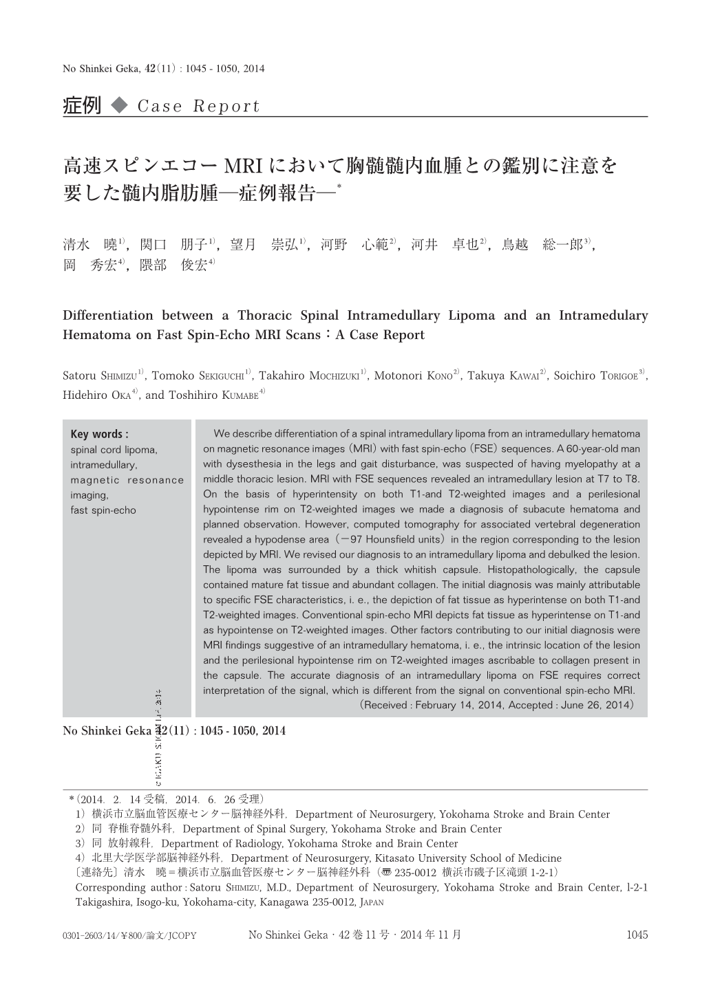
We describe differentiation of a spinal intramedullary lipoma from an intramedullary hematoma on magnetic resonance images(MRI)with fast spin-echo(FSE)sequences. A 60-year-old man with dysesthesia in the legs and gait disturbance, was suspected of having myelopathy at a middle thoracic lesion. MRI with FSE sequences revealed an intramedullary lesion at T7 to T8. On the basis of hyperintensity on both T1-and T2-weighted images and a perilesional hypointense rim on T2-weighted images we made a diagnosis of subacute hematoma and planned observation. However, computed tomography for associated vertebral degeneration revealed a hypodense area(−97 Hounsfield units)in the region corresponding to the lesion depicted by MRI. We revised our diagnosis to an intramedullary lipoma and debulked the lesion. The lipoma was surrounded by a thick whitish capsule. Histopathologically, the capsule contained mature fat tissue and abundant collagen. The initial diagnosis was mainly attributable to specific FSE characteristics, i. e., the depiction of fat tissue as hyperintense on both T1-and T2-weighted images. Conventional spin-echo MRI depicts fat tissue as hyperintense on T1-and as hypointense on T2-weighted images. Other factors contributing to our initial diagnosis were MRI findings suggestive of an intramedullary hematoma, i. e., the intrinsic location of the lesion and the perilesional hypointense rim on T2-weighted images ascribable to collagen present in the capsule. The accurate diagnosis of an intramedullary lipoma on FSE requires correct interpretation of the signal, which is different from the signal on conventional spin-echo MRI.

Copyright © 2014, Igaku-Shoin Ltd. All rights reserved.


