Japanese
English
- 有料閲覧
- Abstract 文献概要
- 1ページ目 Look Inside
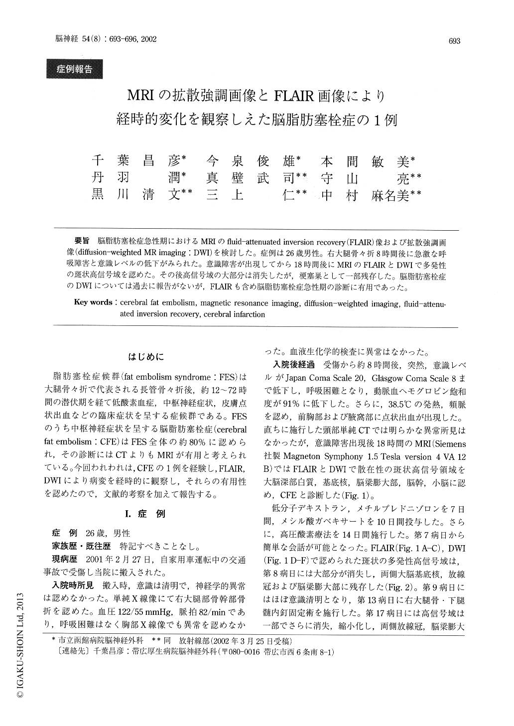
脳脂肪塞栓症急性期におけるMRIのfluid-attenuated inversion recovery(FLAIR)像および拡散強調画像(diffusion-weighted MR imaging:DWI)を検討した。症例は26歳男性。右大腿骨々折8時間後に急激な呼吸障害と意識レベルの低下がみられた。意識障害が出現してから18時間後にMRIのFLAIRとDWIで多発性の斑状高信号域を認めた。その後高信号域の大部分は消失したが,梗塞巣として一部残存した。脳脂肪塞栓症のDWIについては過去に報告がないが,FLAIRも含め脳脂肪塞栓症急性期の診断に有用であった。
Cerebral fat embolism (CFE) is serious complication of a long-bone fracture. We reported magnetic reso-nance (MR) diffusion-weighted (DWI) and fluid at-tenuated inversion recovery (FLAIR) images in a pa-tient suffered with CFE. A 26-year-old man with a right femoral bone fracture lapsed into a semicoma eight hours later. Eighteen hours after the depressed consciousness, DWI and FLAIR images on MR imag-ing showed multiple high-intensity spots in corona ra-diata, basal ganglia, thalamus, corpus callosum, brain stem and cerebellum.

Copyright © 2002, Igaku-Shoin Ltd. All rights reserved.


