Japanese
English
- 有料閲覧
- Abstract 文献概要
- 1ページ目 Look Inside
Ι.はじめに
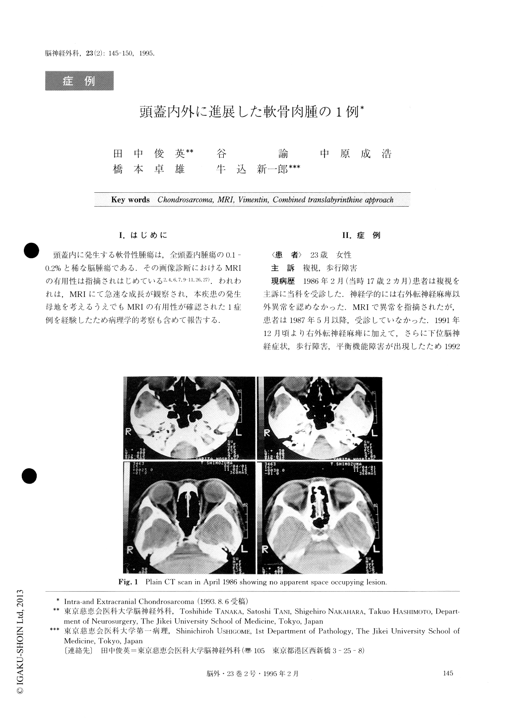
頭蓋内に発生する軟骨性腫瘍は,全頭蓋内腫瘍の0.1—0.2%と稀な脳腫瘍である.その画像診断におけるMRIの有用性は指摘されはじめている2,4,6,7,9-11,26,27).われわれは,MRIにて急速な成長が観察され,本疾患の発生母地を考えるうえでもMRIの有用性が確認された1症例を経験したため病理学的考察も含めて報告する.
A 17-year-old female, who had diplopia and progres-sive gait disturbance, had been diagnosed as having a right parasellar mass lesion in 1986. Initial CT scan failed to show a parasellar space occupying lesion, al-though it was well demonstrated on MRI. No surgery was carried out at that time.
Seven years later, she was hospitalized because of progressive gait disturbance, lower cranial nerves palsy, and cerebellar sign as well as the known right abdu-cence palsy. At this time, CT scan exhibited a huge low density area with a marginal high density area sug-gesting calcification, which extended from the right pa-rasellar area to the ventral portion of the midbrain. She underwent surgery under the diagnosis of cartilaginous tumor or chordoma. The intracranial lesion was re-moved via combined translabyrinthine approach and epidural subtemporal approach. The tumor was hitolo-gically diagnosed as well-differentiated chondrosarco-ma. Electron-microscopy demonstrated tumor cells with features of chondrocytes.
Since the original site of the tumor, strategies for treatment and prognosis are different from each other, distinction between chondrosarcoma and chordoma is important. MRI seemed very useful for making this dis-tinction as a first step.
Copyright © 1995, Igaku-Shoin Ltd. All rights reserved.