Japanese
English
- 有料閲覧
- Abstract 文献概要
- 1ページ目 Look Inside
- 参考文献 Reference
Ⅰ.はじめに
遺伝性出血性毛細血管拡張症(hereditary hemorrhagic telangiectasia:HHT)は常染色体優性の遺伝疾患であり,鼻出血,肺・肝・中枢神経系などの諸臓器の動静脈奇形(arteriovenous malformation:AVM),および顔面・口腔・手指などの毛細血管拡張を特徴とする2,8,14).本邦においても5,000〜8,000人に1人の有病率と考えられ,1万人以上の患者が存在することになるが4,14),決して認知度は高くない.HHTにおいては,全身合併症の除外や血縁者のスクリーニングが必要になり,脳神経外科領域ではAVM症例を診療する場合にはHHTを念頭に置く必要がある.
脳動静脈奇形(脳AVM)はHHTに高率に合併するが,HHTに合併した脳AVMの出血率は孤発AVMに比べて低いとされる1,2,19).今回,出血発症の脳AVMの精査の過程でHHTと診断された症例を経験したため報告する.
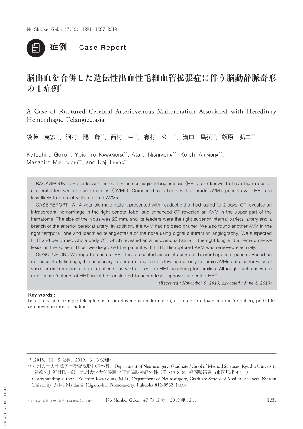
BACKGROUND:Patients with hereditary hemorrhagic telangiectasia(HHT)are known to have high rates of cerebral arteriovenous malformations(AVMs). Compared to patients with sporadic AVMs, patients with HHT are less likely to present with ruptured AVMs.
CASE REPORT:A 14-year-old male patient presented with headache that had lasted for 2 days. CT revealed an intracerebral hemorrhage in the right parietal lobe, and enhanced CT revealed an AVM in the upper part of the hematoma. The size of the nidus was 20 mm, and its feeders were the right superior internal parietal artery and a branch of the anterior cerebral artery. In addition, the AVM had no deep drainer. We also found another AVM in the right temporal lobe and identified telangiectasia of the nose using digital subtraction angiography. We suspected HHT and performed whole body CT, which revealed an arteriovenous fistula in the right lung and a hematoma-like lesion in the spleen. Thus, we diagnosed the patient with HHT. His ruptured AVM was removed electively.
CONCLUSION:We report a case of HHT that presented as an intracerebral hemorrhage in a patient. Based on our case study findings, it is necessary to perform long-term follow-up not only for brain AVMs but also for visceral vascular malformations in such patients, as well as perform HHT screening for families. Although such cases are rare, some features of HHT must be considered to accurately diagnose suspected HHT.

Copyright © 2019, Igaku-Shoin Ltd. All rights reserved.


