Japanese
English
- 有料閲覧
- Abstract 文献概要
- 1ページ目 Look Inside
I.はじめに
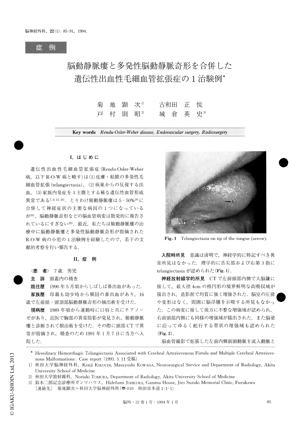
遺伝性出血性毛細血管拡張症(Rendu-Osler-Weber病,以下R-O—W病と略す)は(1)皮膚・粘膜の多発性毛細血管拡張(telangiectasia),(2)病巣からの反復する出血,(3)家族内発症を3主徴とする稀な遺伝性血管形成異常である7,9,13,16).とりわけ肺動静脈瘻は5-50%19)に合併して神経症状の主要な病因の1つになっているが16),脳動静脈奇形などの脳血管病変は散発的に報告されているにすぎない20).最近,私たちは肺動静脈瘻の治療中に脳動静脈瘻と多発性脳動静脈奇形が指摘されたR-O—W病の小児の1治験例を経験したので,若干の文献的考察を行い報告する.
Hereditary hemorrhagic telangiectasia (HHT), or Ren-du-Osler-Weber disease, is an autosomal dominant dis-order characterized by a triad of mucocutaneous and visceral telangiectasia, recurrent epistaxis and familial history. We reported a rare case of HHT associated with pulmonary and cerebral arteriovenous fistulae (AVF) and multiple cerebral arteriovenous malformations (AVM). The roles of multimodality therapies including artificial embolization, feeder clipping and stereotactic radiosurgery for these multiple cerebrovascular dyspla-sia in HHT were discussed.
A 7-year-old boy had presented himself at another hos-pital 2 years previously with cyanosis of the lips and fin-gers on exertion. He was diagnosed as having pulmonary AVF and underwent surgery. His mother had suffered from epistaxis in her adolescence, and was then highly suspected as having HHT. She underwent surgical remo-val of a left fronto-parietal AVM at the age of 16 years. The family history then prompted the patient to have a brain CT done, which eventually demonstrated an abnor-mal enhancing mass at the left frontal region. He was transferred to our service for further evaluation.
Left carotid angiograms demonstrated an AVF sup-plied by a dilated anterior internal frontal artery of theanterior cerebral artery (ACA), draining directly into the vein of the corpus collosum with a large aneurysmal dilatation, and then draining further into the straight sinus via the vein of Galen. In addition, right carotid angiograms revealed three small AVM's fed by the me-dian artery of the corpus callosum, and the middle inter-nal frontal and paracentral arteries of the right ACA, re-spectively. Tracker-18 catheter was placed selectively into the feeder of the AVF and Hilal minicoil was care-fully injected. However, the minicoil failed to remain in the AVF and moved swiftly into the drainer, and even-tually disappeared. It was confirmed that the coil wascaught in the hilum of the left lung. Seven days after this endovascular surgery he underwent a bifrontal cra-niotomy. The AVF was exposed by the interhemispheric approach and a Sugita's miniclip was placed on the feed-er. About one year later the patient was further treated with stereotactic radiosurgery for three AVM's. Total doses ranged from 25 to 31.3 Gy with 80-90% covering isodose. Follow-up angiography done 8 months after radiosurgery confirmed complete obliteration of all the AVM's.

Copyright © 1994, Igaku-Shoin Ltd. All rights reserved.


