Japanese
English
- 有料閲覧
- Abstract 文献概要
- 1ページ目 Look Inside
I.はじめに
近年,本邦においては肺結核による死亡率は減少を続けており11),それに伴って頭蓋内結核腫の頻度も低下している1,10,19).しかし,この結核腫もCT導入後のここ数年間,少数例ではあるが症例報告され9,14,20,27),決して稀な疾患とはいえない.
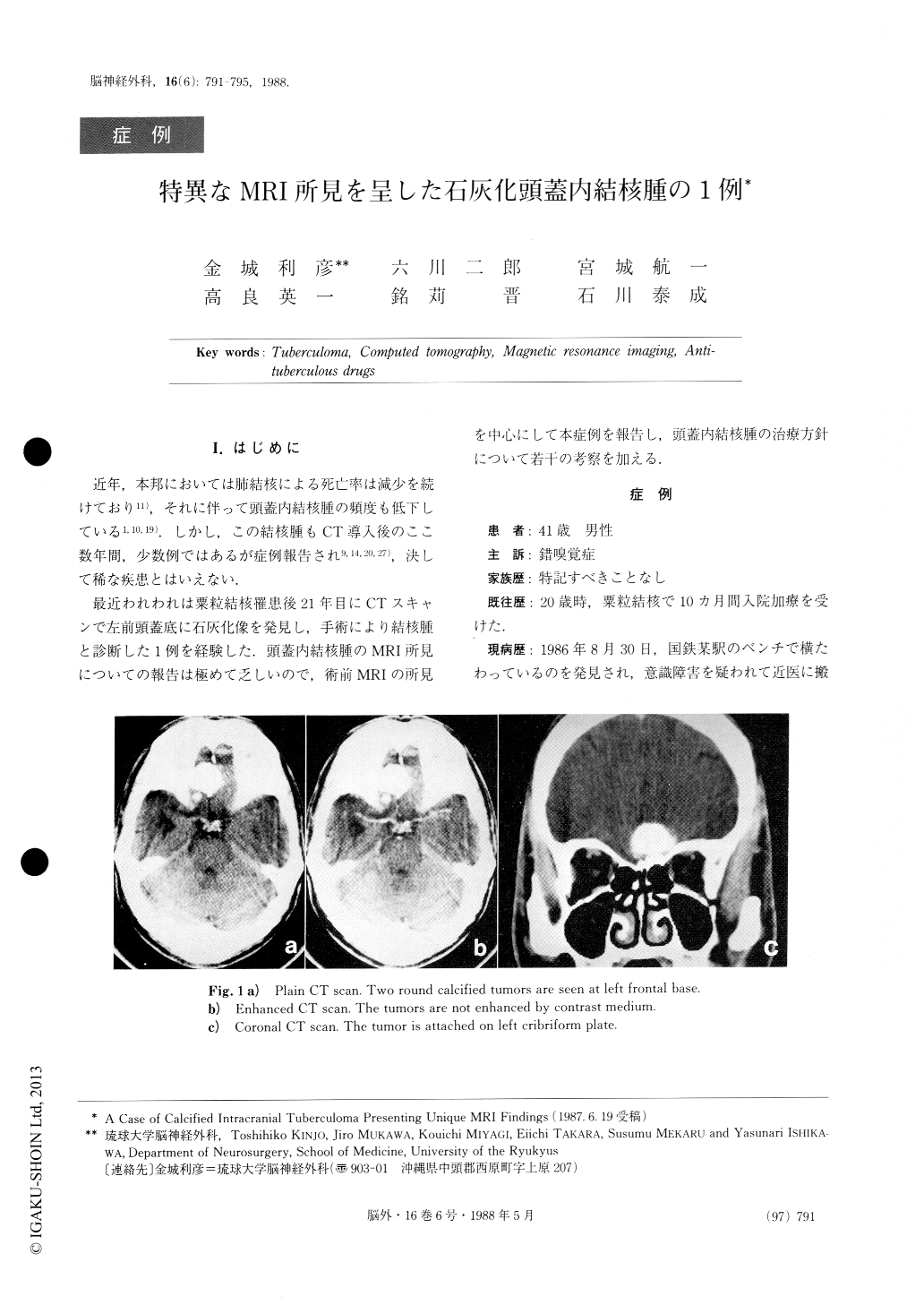
最近われわれは粟粒結核罹患後21年目にCTスキャンで左前頭蓋底に石灰化像を発見し,手術により結核腫と診断した1例を経験した.頭蓋内結核腫のMRI所見についての報告は極めて乏しいので,術前MRIの所見を中心にして本症例を報告し,頭蓋内結核腫の治療方針について若干の考察を加える.
A 41-year-old male patient was admitted in our Ryukyu University Hospital complaining of parosmia. He had a history of miliary tuberculosis 21 years ago. Neurologically he showed left anosmia and hyperreflex-ia of the right upper extremity. Plain skull X-P and CT scan revealed a calcified mass, 25mm in diameter, at the left frontal base. In MRI, the mass showed isoin-tensity using the Ti weighted inversion recovery sequ-ence and heterogenously low intensity using the T, weighted spin echo sequence.
Surgery was performed by bifrontal craniotomy. Then the tumor was removed totally including two coexisting small tumors.

Copyright © 1988, Igaku-Shoin Ltd. All rights reserved.


