Japanese
English
- 有料閲覧
PPV購入案内
1,320円 (1,200円+税10%) こちらは、547ページから550ページの文献です。購入には医書.jp本会員登録が必要です。
会員登録完了後に改めて上記「購入サイトへ」を選択してください。
または「購入サイトへ」選択後に「購入ログイン」、「新規会員登録」の順に進んでください。
会員登録について詳しくはをご確認ください。
- Abstract 文献概要
- 1ページ目 Look Inside
I.はじめに
最近,私どもはsuprasellar germinomaの摘出及び放射線治療後1年で再発が疑われて再開頭し,組織学的に異物性肉芽腫と診断された1例を経験した.脳腫瘍摘出後に発生した異物性肉芽腫の報告は少ない2,17-19).異物性肉芽腫の術前診断について術前MRIも加えて考察し報告する.
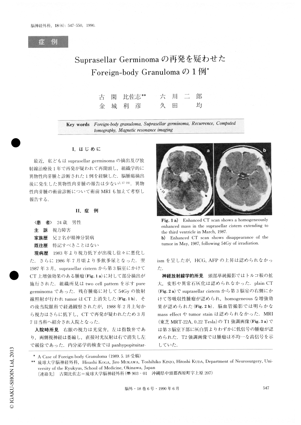
A 24-year-old male patient was admitted to our Ryukyu University Hospital, complaining of visual dis-turbance. He had had partial removal of a suprasellar region tumor in another hospital one year before the admission. Microscopical findings had shown two cell patterns of germinoma in the first operation. Following it, the patient received irradiation with a total close of 54Gy. The tumor completely disappeared after these procedures.
On this admission, plain CT scan revealed an isodensity mass in the suprasellar cistern extending to the right side of the third ventricle, which was en-hanced homogeneously. In MRI, the mass showed low intensity in the T1-weighted inversion recovery sequ-ence, and heterogeneously, high intensity in the T2-weighted spin echo sequence. By bifrontal craniotomy, the tumor was removed. Histologically, it consisted of granuloma containing fine cotton fibers.
MRI findings of intracranial foreign-body granuloma were discussed.
Copyright © 1990, Igaku-Shoin Ltd. All rights reserved.