Japanese
English
- 有料閲覧
PPV購入案内
1,320円 (1,200円+税10%) こちらは、161ページから164ページの文献です。購入には医書.jp本会員登録が必要です。
会員登録完了後に改めて上記「購入サイトへ」を選択してください。
または「購入サイトへ」選択後に「購入ログイン」、「新規会員登録」の順に進んでください。
会員登録について詳しくはをご確認ください。
- Abstract 文献概要
- 1ページ目 Look Inside
I.はじめに
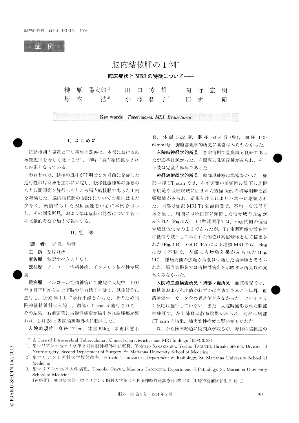
抗結核剤の発達と予防衛生の改善は,本邦における結核罹患率を著しく低下させ9),同時に脳内結核腫もまれな疾患となっている.
われわれは,結核の既往が不明で5カ月前に発症した進行性の片麻痺を主訴に来院し,転移性脳腫瘍の診断のもとに開頭術を施行したところ脳内結核腫であった1例を経験した.脳内結核腫のMRIについての報告は未だ少なく,術前得られたMR画像を中心に本例を呈示し,その画像所見,および臨床症状の特徴について若干の文献的考察を加えて報告する.
A case of intracerebral tuberculoma treated surgically was reported. A 47-year-old man was admitted to our hospital because of progressive left hemiparesis over the previous 5 months. A computerized tomography scan show-ed a well enhanced mass associated with a marked perifocal edema. T1 weighted magnetic resonance imaging (MRI) revealed an isosignal ring around the heterogenous low intensity mass. A low intensity area just interior to this ring was also visualized both in T1 and T2 weighted im-ages. Although the clinical course was unusually long, this was diagnosed as a metastatic brain tumor. He underwent a right frontal craniotomy and a well circum-scribed, yellowish, firm mass was totally extirpated. Pathohistologically, this mass was considered to be a tuberculoma though the tuberculous bacilli could not be identified in Ziehl-Neelsen staining. His hemiplegia im-proved much and his ambulation was restored.
Since tuberclomas are quite rare in developed coun-tries, the diagnosis of intracerebral tuberculomas would be extremely difficult unless tuberculosis was verified in some other organs. The auxiliary examinations even by using MRI have often given little information which would assist diagnossis. However, based on patholo-gical findings, the ring appearance and low intensity area medial to the ring in the outer part of the tuberculoma shown in MRI of our patient seemed to represent a chro-nic granulomatous inflammation and gave a clue to rule out the suspicion of metastatic brain tumors. To make a correct diagnosis of intracerebral tuberculomas, multidis-ciplinary consideration is mandatory.
Copyright © 1994, Igaku-Shoin Ltd. All rights reserved.