Japanese
English
- 有料閲覧
PPV購入案内
1,320円 (1,200円+税10%) こちらは、1167ページから1170ページの文献です。購入には医書.jp本会員登録が必要です。
会員登録完了後に改めて上記「購入サイトへ」を選択してください。
または「購入サイトへ」選択後に「購入ログイン」、「新規会員登録」の順に進んでください。
会員登録について詳しくはをご確認ください。
- Abstract 文献概要
- 1ページ目 Look Inside
I.はじめに
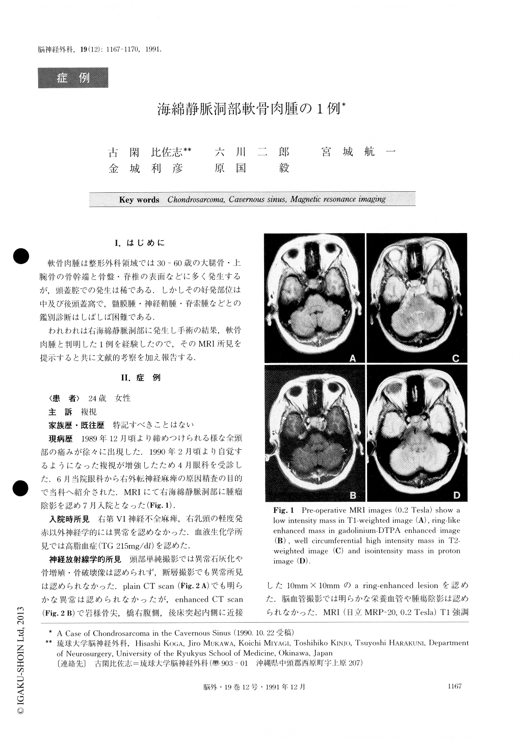
軟骨肉腫は整形外科領域では30-60歳の大腿骨・上腕骨の骨幹端と骨盤・脊椎の表面などに多く発生するが,頭蓋腔での発生は稀である.しかしその好発部位は中及び後頭蓋窩で,髄膜腫・神経鞘腫・脊索腫などとの鑑別診断はしばしば困難である.
われわれは右海綿静脈洞部に発生し手術の結果,軟骨肉腫と判明した1例を経験したので,そのMRI所見を提示すると共に文献的考察を加え報告する.
A 24-year-old female patient complained of headache and right abducens nerve paralysis. No abnormality was found in plain CT scan, but a ring-like enhanced mass was disclosed behind the right posterior clinoid process in enhanced CT scan. MRI revealed a low in-tensity mass in Tl-weighted image and a ring-like en-hanced mass in gadolinium-DTPA enhanced image. It was a circumferential high intensity mass in T2-weighted image and an isointensity mass in proton im-age. Cerebral angiography indicated that it was avascu-lar. After discharge he was in good health, and had had some follow up, CT were normal except for the hematoma cavity.
Preoperative diagnosis was trigeminal neurinoma or petroclival meningioma. The tumor was removed almost completely by orbi-tozygomatic infratemporal approach. Histologically, it was low grade chondrosarcoma. Postoperatively, neith-er radiation therapy nor chemotherapy was added. Differential diagnosis and treatment was discussed. It was suggested that MRI was the most useful diagnostic tool to distinguish chondrosarcoma from other skull base tumors.
Copyright © 1991, Igaku-Shoin Ltd. All rights reserved.