Japanese
English
症例
異なる組織像を示したMulticentric gliomaの1剖検例
An Autopsy Case of Multicentric Glioma of Multiple Histopathology
中瀬 裕之
1
,
久永 学
1
,
岩永 秀昭
1
Hiroyuki NAKASE
1
,
Manabu HISANAGA
1
,
Hideaki IWANAGA
1
1国立泉北病院脳神経外科
1Department of Neurosurgery, National Senboku Hospital
キーワード:
Multicentric glioma
,
Glioma
,
Computed tomography
Keyword:
Multicentric glioma
,
Glioma
,
Computed tomography
pp.1073-1077
発行日 1987年10月10日
Published Date 1987/10/10
DOI https://doi.org/10.11477/mf.1436202481
- 有料閲覧
- Abstract 文献概要
- 1ページ目 Look Inside
I.はじめに
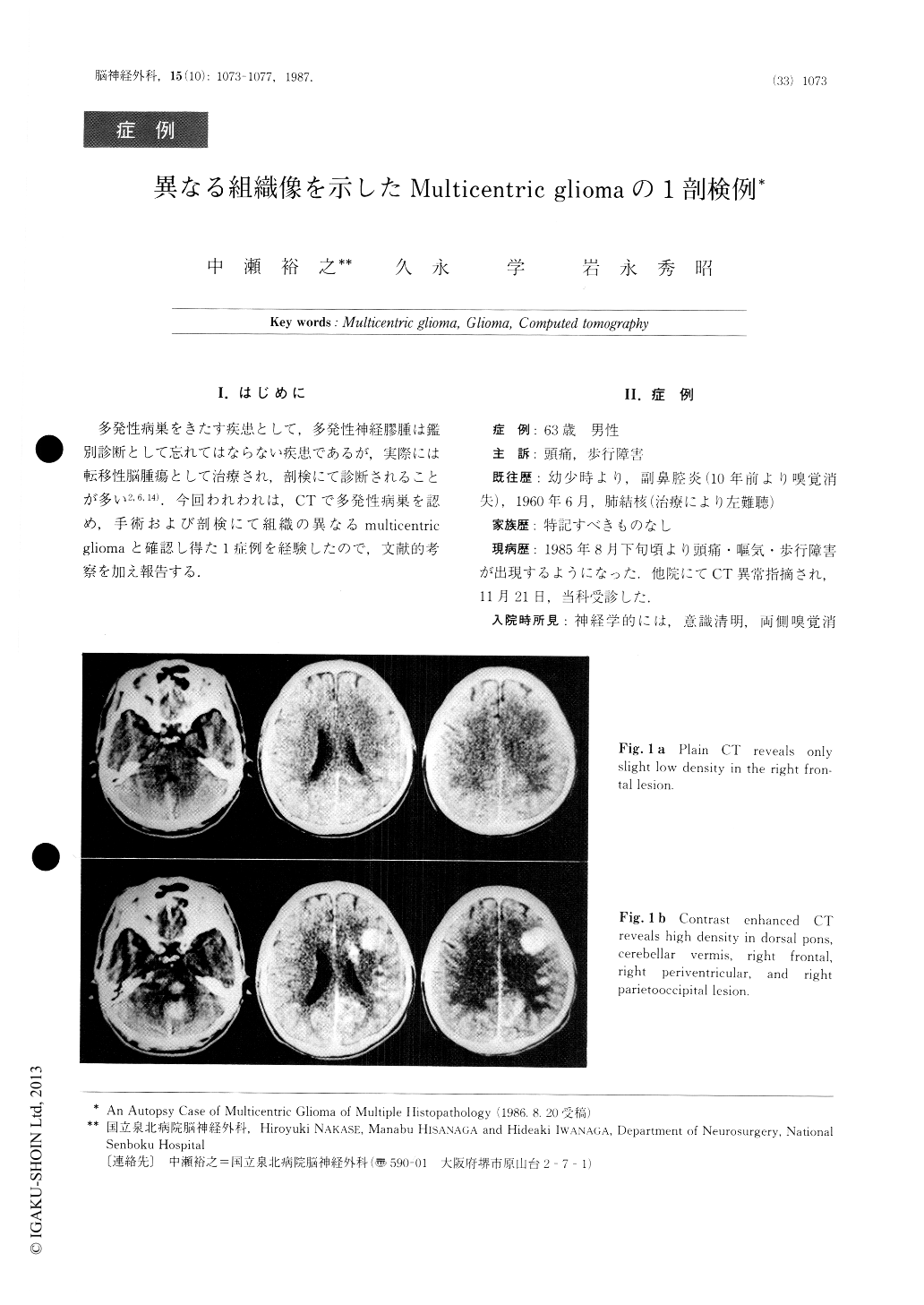
多発性病巣をきたす疾患として,多発性神経膠腫は鑑別診断として忘れてはならない疾患であるが,実際には転移性脳腫瘍として治療され,剖検にて診断されることが多い2,6,14).今回われわれは,CTで多発性病巣を認め,手術および剖検にて組織の異なるmulticentricgliomaと確認し得た1症例を経験したので,文献的考察を加え報告する.
An autopsy case is described of an 66-year-old man with multicentric glioma of multiple histopathology, i.e. protoplasmic astrocytoma and glioblastoma.
Enhanced CT scan revealed three separate lesions in the right cerebral hemisphere, pons, and cerebellar vermis. Initial diagnosis by CT included metastatic and primary brain tumor, multiple abscess, fungal infec-tion, parasites, tuberculoma, and so on. Biopsy of the right frontal mass revealed astrocytoma grade-2.

Copyright © 1987, Igaku-Shoin Ltd. All rights reserved.


