Japanese
English
- 有料閲覧
- Abstract 文献概要
- 1ページ目 Look Inside
I.はじめに
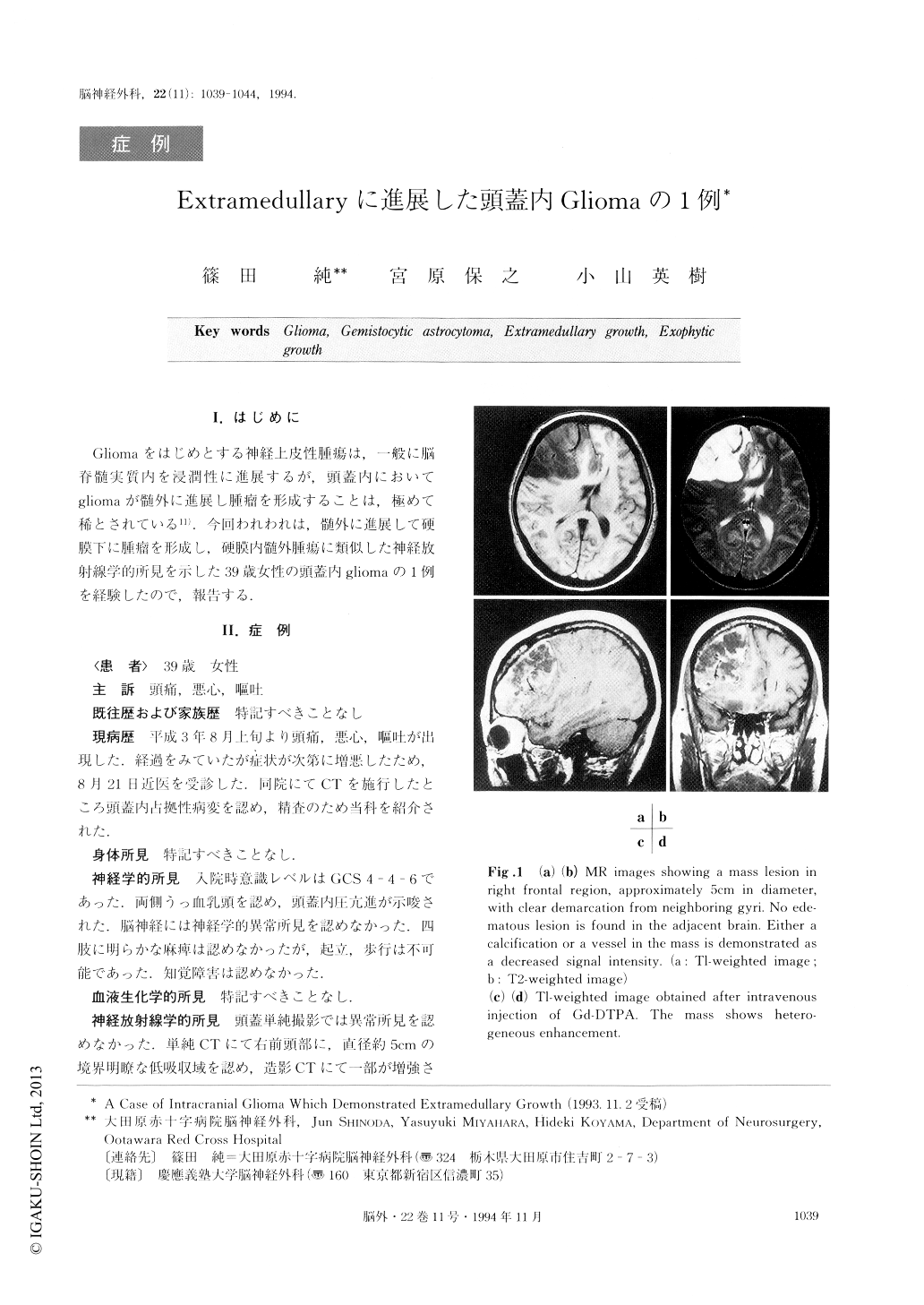
Gliomaをはじめとする神経上皮性腫瘍は,一般に脳脊髄実質内を浸潤性に進展するが,頭蓋内においてgliomaが髄外に進展し腫瘤を形成することは,極めて稀とされている11).今回われわれは,髄外に進展して硬膜下に腫瘤を形成し,硬膜内髄外腫瘍に類似した神経放射線学的所見を示した39歳女性の頭蓋内gliomaの1例を経験したので,報告する.
An unusual case of an intracranial glioma which de-monstrated extramedullary growth is reported.
The patient was a 39-year-old woman who had ex-perienced headache, nausea and vomiting for about 1 month. On admission she showed slight disturbance of consciousness and bilateral papilloedema. CT scan and MR imaging disclosed a mass approximately 5 cm in diameter in the right frontal region, with clear demarca-tion from the neighboring gyri. Right external carotid angiogram revealed A-V shunts in the mass, but by right internal carotid angiogram, no abnormal findings were disclosed except for the deviation of normal intra-cranial vessels due to the existence of the mass. There-fore, a preoperative diagnosis of extramedullary tumor such as meningioma and epidermoid was made.
Right frontal craniotomy was performed, and thetumor was proven to exist in subdural space. The boundary of the tumor to the brain surface was distinct except for one part. Histopathologically, the tumor cells had abundant eosinophillic cytoplasms, with eccentric-distribution of their nuclei. Furthermore, they were positive for staining for GFAP and S-100 protein. Therefore, a final diagnosis of gemistocytic astrocyto-ma was made.
Reviewing some references the authors discuss here the form of development and progression of intracranial gliomas which demonstrate extramedullary growth such as this case.

Copyright © 1994, Igaku-Shoin Ltd. All rights reserved.


