Japanese
English
- 有料閲覧
- Abstract 文献概要
- 1ページ目 Look Inside
I.はじめに
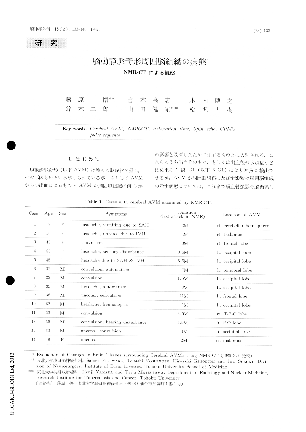
脳動静脈奇形(以下AVM)は種々の脳症状を呈し,その原因もいろいろ挙げられているが,上としてAVMからの出血.によるものとAVMが周囲脳組織に何らかの影響を及ぼしたために生ずるものとに大別される.これらのうち出血そのもの,もしくは出血後の水頭症などは従来のX線CT(以下X-CT)により容易に検出できるが,AVMが周囲脳組織に及ぼす影響や周囲脳組織の示す病態については,これ虫で脳血管撮影や脳循環などの立場から種々の議論がなされてはいるものの三次元的把握は困難であった.そこでわれわれはこのAVM周囲脳組織の病態解明の一助としてNMR-CTを用い,AVMを含めた周囲脳組織のイメージング,T1・T2計算画像による観察および関心領域(以下ROI)のT1・T2緩和時間値の測定を試みたので報告する.
Patients with cerebral arteriovenous malformation (AVM) show various clinical symptoms, but they can be divided into two groups; one resulting from rupture of AVM and another derived from chronic ischemia in surrounding tissues of AVM.
Intracerebral or suharachnoid hemorrhages due to rupture of AVM can be detected by X-ray CT scan, however, it is difficult to obtain three dimensional image of changes in the surrounding area of AVM that has never experienced hemorrhagic attacks.

Copyright © 1987, Igaku-Shoin Ltd. All rights reserved.


