Japanese
English
- 有料閲覧
- Abstract 文献概要
- 1ページ目 Look Inside
I.はじめに
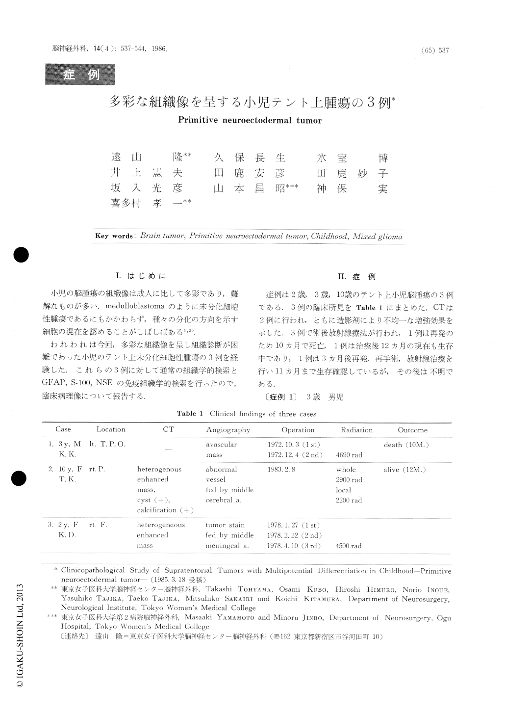
小児の脳腫瘍の組織像は成人に比して多彩であり,難解なものが多い.medulloblastomaのように未分化細胞性腫瘍であるにもかかわらず,種々の分化の方向を示す細胞の混在を認めることがしばしばある1,2).
われわれは今回,多彩な組織像を呈し組織診断が困難であった小児のテント上未分化細胞性腫瘍の3例を経験した.これらの3例に対して通常の組織学的検索とGFAP,S−100,NSEの免疫組織学的検索を行ったので,臨床病理像について報告する.
Three cases of supratentorial tumor in childhood were studied clinico-pathologically in an attempt to clarify its histological character.
Case 1: A 3-year-old boy. Carotid angiogram re-vealed avascular lesion in the left parietal lobe. Twice operations and radiotherapy were performed. Ten months after the second operation, he (lied. Surgical specimen at the first operation was composed mainly of round tumor cells. The tumor tissue contained many collagen fibers. At the periphery of this tissue, medulloblastomatous areas consisting of closely ag-gregated hyperchromatic small round cells were found.

Copyright © 1986, Igaku-Shoin Ltd. All rights reserved.


