Japanese
English
- 有料閲覧
- Abstract 文献概要
- 1ページ目 Look Inside
I.はじめに
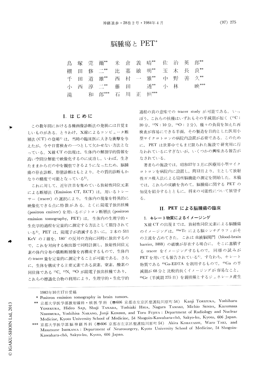
この数年間における各種画像診断法の発展には目覚ましいものがある。とりわけ,X線によるコンピュータ断層法(CT)の登場1)は,当時の臨床医に大きな衝撃を与えたが,今や日常検査の一つとして欠かせない方法となっている。X線CTの出現は,生体内の解剖学的情報を高い空間分解能で映像化するのに成功し,いわば,生きたままからだの中を観察できるようになったため,脳腫瘍の存在診断,形態診断はもとより,その質的診断もかなりの精度で可能となっている2)。
これに対して,近年注目を集めている放射性同位元素による断層法(Emission CT,ECT)は,用いるトレーサー(tracer)の選択により,生体内の現象を特異的に映像化できる点に特徴がある。とくに陽電子放出核種(positron emitter)を用いるポジトロン断層法(positronemission tomography,PET)は,生体内の生理学的・生化学的過程を定量的に測定する方法として期待されている3)。PETは,陽電子が消滅するさいに,2本の511KeVのγ線を,180°の反対の方向に同時に放出するので,これを対向する検出器で同時計測し,放射性同位元素の体内分布の横断断層像を再構成するもので,生体内のtracer量を定量的に測定することが可能である。
Abstract
Positron emission tomography (PET) has been exploring pathophysiology and biochemistry in normal and diseased states. Major advantages of PET are i) intrinsic quantitative capability, ii) availability to label substrates of physiological interest and iii) use of ultrashort lived tracers which permits repeat studies in the same subjects with various interventions. This paper describes the current status and the future directions of PET studies in brain tumors.
Brain scintigraphy with 99mTc-pertechnetate had been widely used in clinical nuclear medicine until the introduction of X-ray computed tomo-graphy.

Copyright © 1984, Igaku-Shoin Ltd. All rights reserved.


