Japanese
English
- 有料閲覧
- Abstract 文献概要
- 1ページ目 Look Inside
I.はじめに
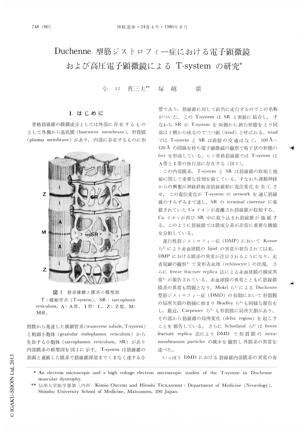
骨格筋線維の膜構成分としては外部に存在するものとして外側から基底膜(basement membrane),形質膜(plasma membrane)があり,内部に存在するものに形質膜から発達した横細管系(transverse tubule,T-system)と粗面小胞体(granular endoplasmic reticulum)から生長する小胞体(sarcoplasmic reticulum,SR)があり内部膜系の模型図を図1に示す。T-systemは筋線維の表面と連続した膜系で筋線維深部までくまなく達する小管であり,筋線維に対して直角に走行するのでこの名称がついた。このT-systemはSRと密接に結合し,すなわちSRがT-systemを両側から挾む形態をとり同部は3層から成るので三つ組(triad)と呼ばれる。triadではT-systemとSRは直接の交通はなく,100Å〜120Åの間隔を持ち電子顕微鏡の観察で格子状の形態のfeetを形成している。ヒト骨格筋線維ではT-systemはA帯とI帯の移行部に存在する(図1)。
この内部膜系,T-systemとSRは筋線維の収縮と弛緩に関して重要な役割を演じている。
We reported the changes of the T-system and sarcoplasmic reticulum in Duchenne muscular dystrophy. The purpose of this paper is to check the T-system and sarcoplasmic reticulum in Duchenne muscular dystrophy using lanthanum by high voltage electron microscopy and to compare with the abnormal findings observed by an electron microsopic study.
Muscle biopsies from 7 patients with Duchenne muscular dystrophy and of 5 normal controls were studied. Biopsies which were taken under local anesthesia from the quadriceps femoris or brachial biceps muscle, were fixed for 20 min in 3% glutaraldehyde.

Copyright © 1980, Igaku-Shoin Ltd. All rights reserved.


