Japanese
English
- 有料閲覧
- Abstract 文献概要
- 1ページ目 Look Inside
はじめに
1966年Tanaghoら1)により報告された下部尿路の形態を把える画像診断法と尿水力学的検査法との同時記録および表示方法が,下部尿路機能の把握に有用とされ,各施設に普及しつつある2〜5)。この方法の臨床的に有用な点としては,1)カテーテル,電極などの尿水力学的検査法の測定器具が正確な部位に留置されていることを確認しながら検査をできる,2)尿水力学的検査法のみでは把えることが難しい症例における尿道の部位別動態をも正確に観察することができる,などが挙げられる。
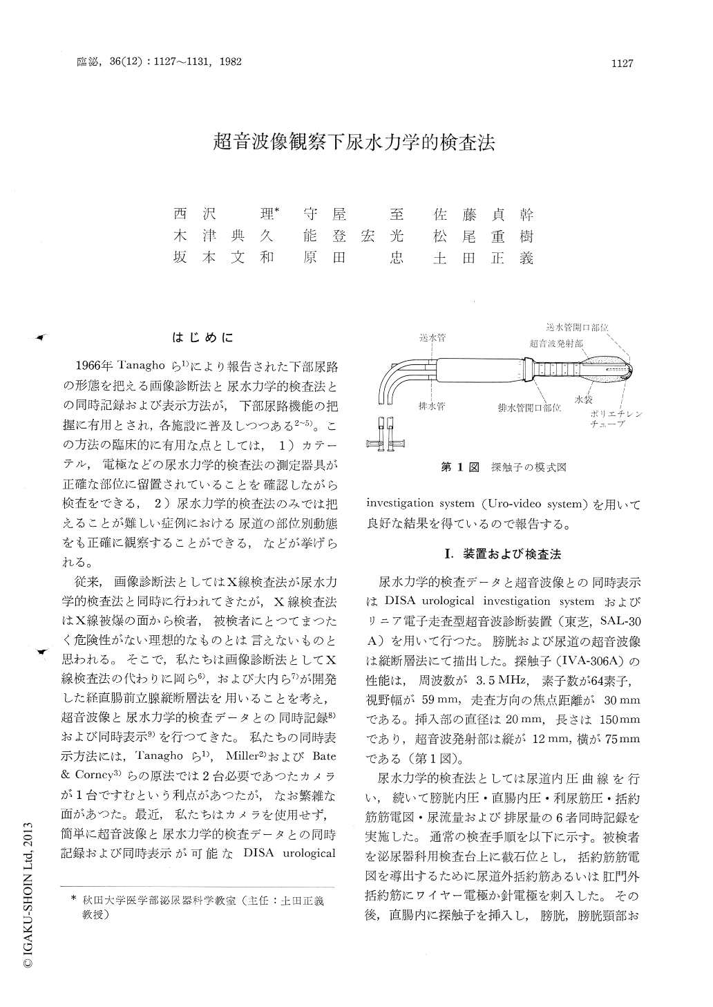
従来,画像診断法としてはX線検査法が尿水力学的検査法と同時に行われてきたが,X線検査法はX線被爆の面から検者,被検者にとつてまつたく危険性がない理想的なものとは言えないものと思われる。そこで,私たちは画像診断法としてX線検査法の代わりに岡ら6),および大内ら7)が開発した経直腸前立腺縦断層法を用いることを考え,超音波像と尿水力学的検査データとの同時記録8)および同時表示9)を行つてきた。
Recently, We have studied the function of the lower urinary tract by the combined ultrasonotomo-graphic and urodynamic monitoring. Transrectal longitudinal ultrasonotomography of the bladder and urethra by electronic linear scanning in real time was performed on sonolayer (Toshiba, SAL-30A). Uro-dynamic examination was performed on the DISA urological investigation system (Uro-Video System) capable of the recording simultaneously external sphincter EMG, total bladder pressure, the rectal pressure, subtracted bladder pressure, voided volume and urine flow rate.

Copyright © 1982, Igaku-Shoin Ltd. All rights reserved.


