Japanese
English
- 有料閲覧
- Abstract 文献概要
- 1ページ目 Look Inside
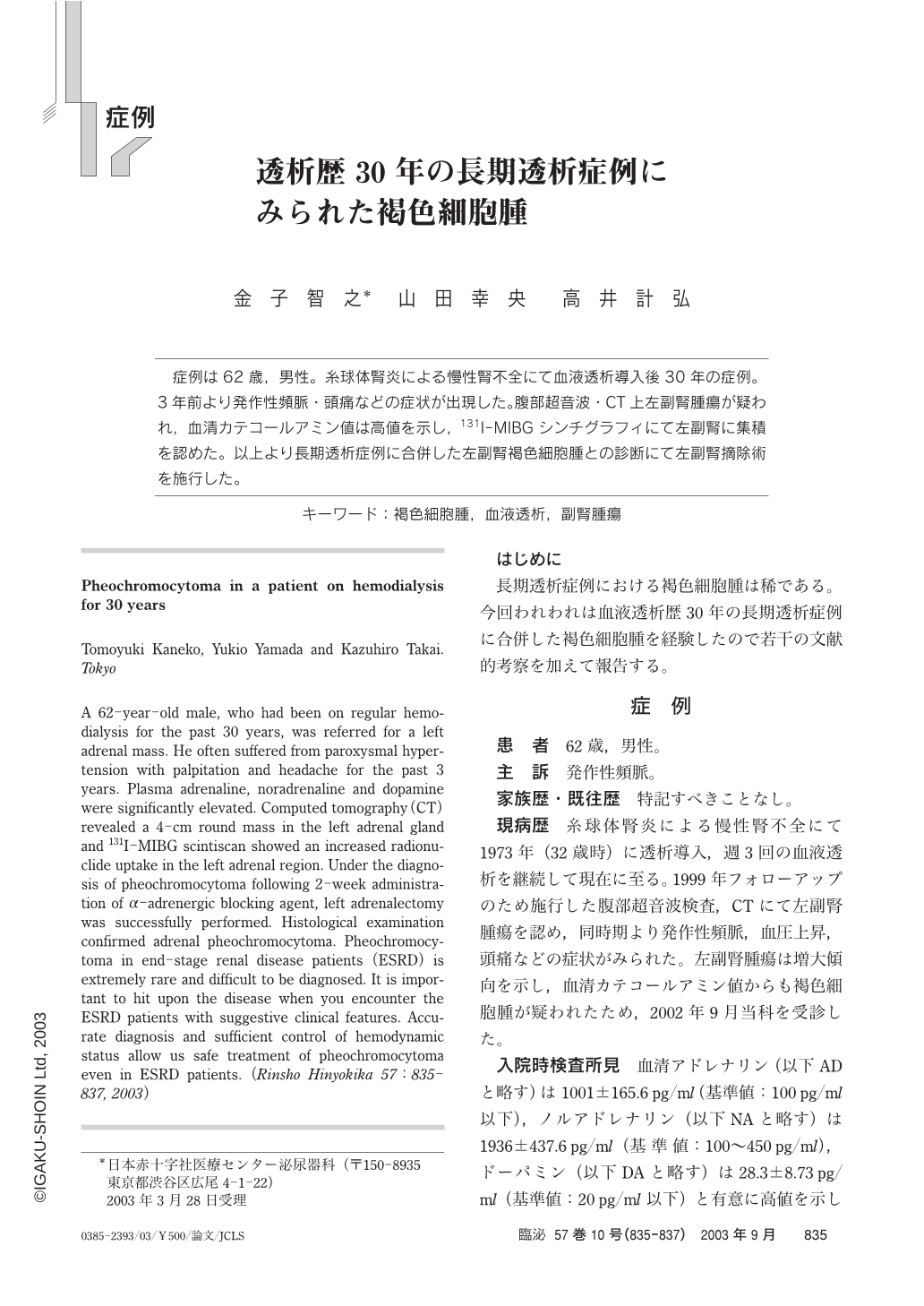
症例は62歳,男性。糸球体腎炎による慢性腎不全にて血液透析導入後30年の症例。3年前より発作性頻脈・頭痛などの症状が出現した。腹部超音波・CT上左副腎腫瘍が疑われ,血清カテコールアミン値は高値を示し,131I-MIBGシンチグラフィにて左副腎に集積を認めた。以上より長期透析症例に合併した左副腎褐色細胞腫との診断にて左副腎摘除術を施行した。
A 62-year-old male,who had been on regular hemodialysis for the past 30 years,was referred for a left adrenal mass. He often suffered from paroxysmal hypertension with palpitation and headache for the past 3 years. Plasma adrenaline,noradrenaline and dopamine were significantly elevated. Computed tomography(CT)revealed a 4-cm round mass in the left adrenal gland and131I-MIBG scintiscan showed an increased radionuclide uptake in the left adrenal region. Under the diagnosis of pheochromocytoma following 2-week administration ofα-adrenergic blocking agent,left adrenalectomy was successfully performed. Histological examination confirmed adrenal pheochromocytoma. Pheochromocytoma in end-stage renal disease patients(ESRD)is extremely rare and difficult to be diagnosed. It is important to hit upon the disease when you encounter the ESRD patients with suggestive clinical features. Accurate diagnosis and sufficient control of hemodynamic status allow us safe treatment of pheochromocytoma even in ESRD patients.

Copyright © 2003, Igaku-Shoin Ltd. All rights reserved.


