Japanese
English
- 有料閲覧
- Abstract 文献概要
- 1ページ目 Look Inside
はじめに
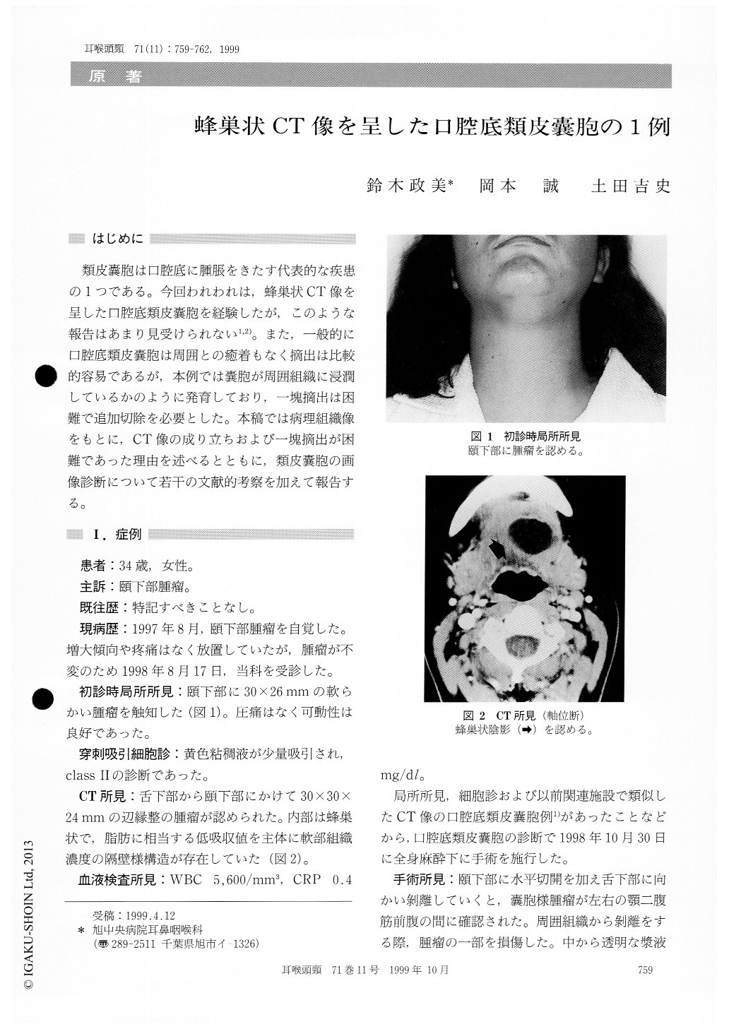
類皮嚢胞は口腔底に腫脹をきたす代表的な疾患の1つである。今回われわれは,蜂巣状CT像を呈した口腔底類皮嚢胞を経験したが,このような報告はあまり見受けられない1,2)。また,一般的に口腔底類皮嚢胞は周囲との癒着もなく摘出は比較的容易であるが,本例では嚢胞が周囲組織に浸潤しているかのように発育しており,一塊摘出は困難で追加切除を必要とした。本稿では病理組織像をもとに,CT像の成り立ちおよび一塊摘出が困難であった理由を述べるとともに,類皮嚢胞の画像診断について若干の文献的考察を加えて報告する。
A 32-year-old femle complained of a submental mass. CT showed a appearance like honeycomb.The cellar area revealed a low density that suggest-ed of fat. The pathological diagnosis was dermoid cyst which contained skin appendages such as seba-ceous glands, hair follicles and others. The cyst filled with pasty yellow material and few liquid. It was thought that fatty small balls and few liquid were mixed in the cyst. The cyst was not removed en block. Pathological specimen showed that the cyst wall was ruptured and there was inflammatory changes in peripheral tissues.

Copyright © 1999, Igaku-Shoin Ltd. All rights reserved.


