Japanese
English
- 有料閲覧
- Abstract 文献概要
- 1ページ目 Look Inside
はじめに
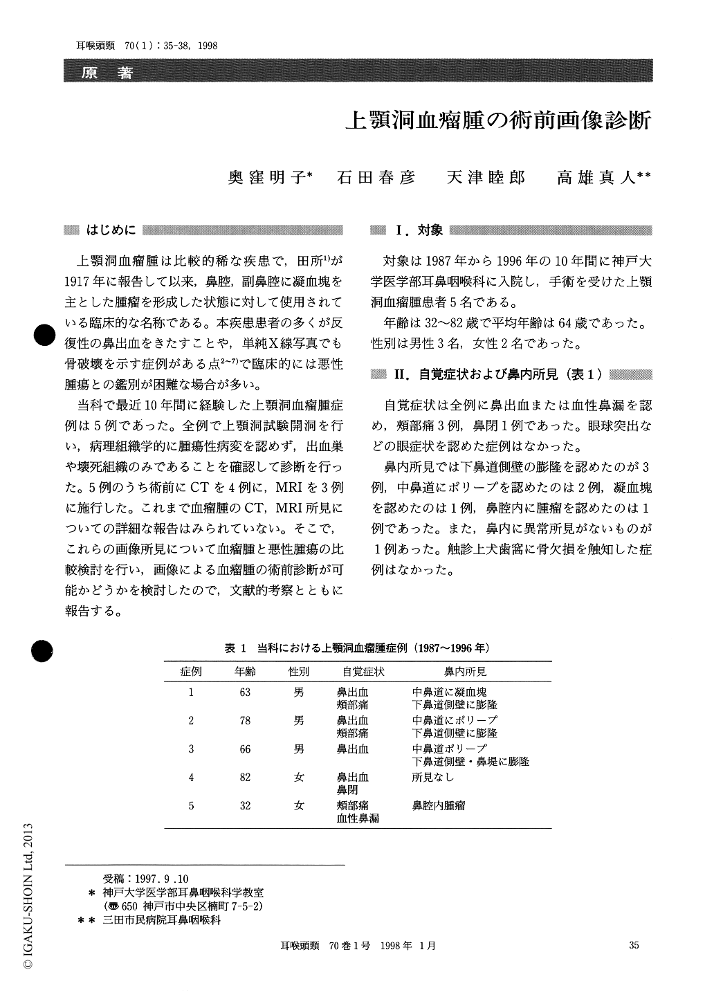
上顎洞血瘤腫は比較的稀な疾患で,田所1)が1917年に報告して以来,鼻腔,副鼻腔に凝血塊を主とした腫瘤を形成した状態に対して使用されている臨床的な名称である。本疾患患者の多くが反復性の鼻出血をきたすことや,単純X線写真でも骨破壊を示す症例がある点2〜7)で臨床的には悪性腫瘍との鑑別が困難な場合が多い。
当科で最近10年間に経験した上顎洞血瘤腫症例は5例であった。全例で上顎洞試験開洞を行い,病理組織学的に腫瘍性病変を認めず,出血巣や壊死組織のみであることを確認して診断を行った。5例のうち術前にCTを4例に,MRIを3例に施行した。これまで血瘤腫のCT,MRI所見についての詳細な報告はみられていない。そこで,これらの画像所見について血瘤腫と悪性腫瘍の比較検討を行い,画像による血瘤腫の術前診断が可能かどうかを検討したので,文献的考察とともに報告する。
We studied five patients with so-called “Blut-beule” at Kobe University Hospital between 1987 and 1996.
CT scans demonstrated unilateral homogeneous maxillary opacification with bony destruction. The mass was well-demarcated from the surrounding tissue. These findings suggested its expansive growth.
In T1-weighted MRI images, isointensity erea coexisted with hyperintensity erea. In T2-weighted MRI images, hyperintensity was found in hypointen-sity erea. These images were clearly demarcated on both Tl-and T2-weighted images campatible with so-called “Blutbeule”.

Copyright © 1998, Igaku-Shoin Ltd. All rights reserved.


