Japanese
English
- 有料閲覧
- Abstract 文献概要
- 1ページ目 Look Inside
緒言
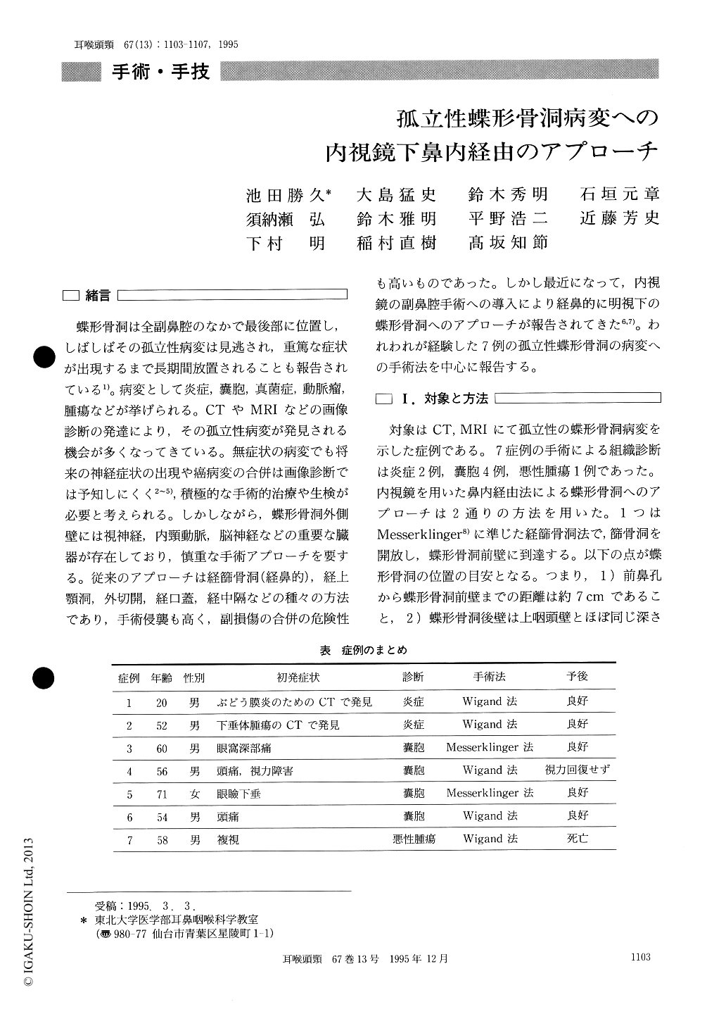
蝶形骨洞は全副鼻腔のなかで最後部に位置し,しばしばその孤立性病変は見逃され,重篤な症状が出現するまで長期間放置されることも報告されている1)。病変として炎症,嚢胞,真菌症,動脈瘤,腫瘍などが挙げられる。CTやMRIなどの画像診断の発達により,その孤立性病変が発見される機会が多くなってきている。無症状の病変でも将来の神経症状の出現や癌病変の合併は画像診断では予知しにくく2〜5),積極的な手術的治療や生検が必要と考えられる。しかしながら,蝶形骨洞外側壁には視神経,内頸動脈,脳神経などの重要な臓器が存在しており,慎重な手術アプローチを要する。従来のアプローチは経篩骨洞(経鼻的),経上顎洞,外切開,経口蓋,経中隔などの種々の方法であり,手術侵襲も高く,副損傷の合併の危険性も高いものであった。しかし最近になって,内視鏡の副鼻腔手術への導入により経鼻的に明視下の蝶形骨洞へのアプローチが報告されてきた6,7)。われわれが経験した7例の孤立性蝶形骨洞の病変への手術法を中心に報告する。
Seven cases with solitary sphenoid sinus lesions diagnosed as inflammation, mucocele or carcinoma underwent endoscopic endonasal sinus surgery. Two surgical approaches to the sphenoid sinus were employed through the ethmoid sinus and the supe-rior meatus. Precise diagnosis and anatomical struc-ture of the sphenoid sinus lesions were achieved with the use of CT scan and MRI. It should be emphasized that the anterior bony wall in the infe-rior/medial quadrant is safely entered due to the absence of the important underlying structures. Endoscopic approach to the sphenoid sinus is benefi-cial with excellent visualization and exposure, less surgical stress on patients, and minimal blood loss and operation time.

Copyright © 1995, Igaku-Shoin Ltd. All rights reserved.


