Japanese
English
原著
鼻茸を伴う鼻・副鼻腔病変のCTによる評価
CT Findings of Nasal Cavity and Paranasal Sinuses in Nasal Polyposis
真崎 正美
1
,
橘 敏郎
2
,
藤原 朋樹
1
,
米本 友明
1
Masami Masaki
1
1東京共済病院耳鼻咽喉科
2聖路加国際病院耳鼻咽喉科
1Department of Otolaryngology, Tokyo Kyosai Hospital
pp.996-998
発行日 1994年11月20日
Published Date 1994/11/20
DOI https://doi.org/10.11477/mf.1411901045
- 有料閲覧
- Abstract 文献概要
- 1ページ目 Look Inside
はじめに
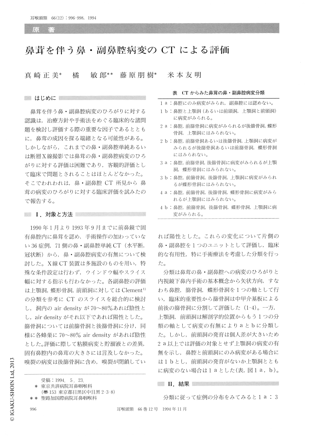
鼻茸を伴う鼻・副鼻腔病変のひろがりに対する認識は,治療方針や手術法をめぐる臨床的な諸問題を検討し評価する際の重要な因子であるとともに,鼻茸の成因を探る端緒となる可能性がある。しかしながら,これまでの鼻・副鼻腔単純あるいは断層X線撮影では鼻茸の鼻・副鼻腔病変のひろがりに対する評価は困難であり,客観的評価として臨床で問題とされることはほとんどなかった。そこでわれわれは,鼻・副鼻腔CT所見から鼻茸の病変のひろがりに対する臨床評価を試みたので報告する。
CT findings of the nasal cavity and paranasal sinuses in nasal polyposis was classified into eight types according to occupying region. The incidence of the nasal cavity involvement was 4.2%. The most nasal polyps showed the pathological changes of the paranasal sinuses, particulary around the eth-moidal sinuses.

Copyright © 1994, Igaku-Shoin Ltd. All rights reserved.


