Japanese
English
- 有料閲覧
- Abstract 文献概要
- 1ページ目 Look Inside
はじめに
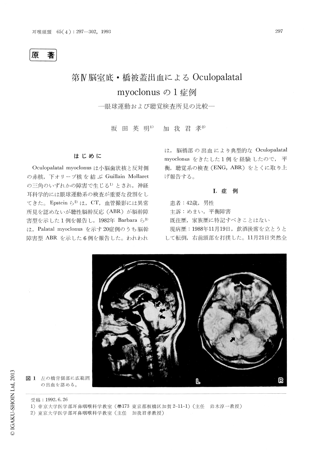
Oculopalatal myoclonusは小脳歯状核と反対側の赤核,下オリーブ核を結ぶGuillain Mollaretの三角のいずれかの障害で生じる1)とされ,神経耳科学的には眼球運動系の検査が重要な役割をしてきた。Epsteinら2)は,CT,血管撮影には異常所見を認めないが聴性脳幹反応(ABR)が脳幹障害型を示した1例を報告し,1982年Barbaraら3)は,Palatal myoclonusを示す20症例のうち脳幹障害型ABRを示した6例を報告した。われわれは,脳橋部の出血により典型的なOculopalatal myoclonusをきたした1例を経験したので,平衡,聴覚系の検査(ENG,ABR)をとくに取り上げ報告する。
We reported a case, 42 years old male, who manifestied ocular, palatal, laryngeal and phar-yngeal myoclonus. A hemorrhagic lesion was demonstrated between the left bottom of the fourth ventricle and the pontine tegmentum.
Eye tracking pattern was ataxic, and optokinctic pattern test indicated cerebellum-brainstem dis-orders. No caloric response was obtained from the left ear. Pure tone audiometry was normal. ABR displayed remarkable prolonged Ⅲ-Ⅴ wave interval at the left. Monosylable discrimination test showed the right ear disadvantage. The above findings seemed to he caused by the left dorsal brainstem lesion composing left central tegmental tract, vestibular nuclei and nucleus of the lateral lemniscus which are called the triangle of Guillain Mollarct. It is noted that the triangle of Guillain Mollaret must be present near auditory brainstem pathway.

Copyright © 1993, Igaku-Shoin Ltd. All rights reserved.


