Japanese
English
- 有料閲覧
- Abstract 文献概要
- 1ページ目 Look Inside
はじめに
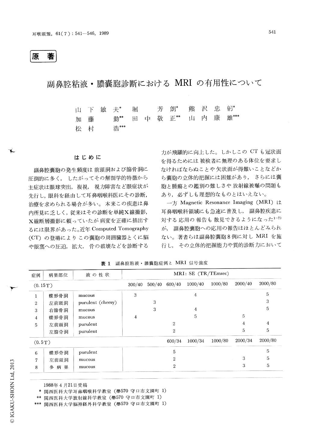
副鼻腔嚢胞の発生頻度は前頭洞および節骨洞に圧倒的に多く,したがってその解剖学的特徴から主症状は眼球突出,複視,視力障害など眼症状が先行し,眼科を経由して耳鼻咽喉科医にその診断,治療を求められる場合が多い。本来この疾患は鼻内所見に乏しく,従来はその診断を単純X線撮影,X線断層撮影に頼っていたが病変を正確に描出するには限界があった。近年Computed Tomography(CT)の登場によりこの嚢胞の周囲臓器とくに脳や眼窩への圧迫,拡大,骨の破壊などを診断する力が飛躍的に向上した。しかしこのCTも冠状面を得るためには被検者に無理のある体位を要求しなければならぬことや矢状面が得難いことなどから嚢胞の立体的把握には困難があり,さらには嚢胞と腫瘍との鑑別の難しさや放射線被曝の問題もあり,必ずしも理想的なものとはいえない。
一方Magnetic Resonance Imaging (MRI)は耳鼻咽喉科領域にも急速に普及し,副鼻腔疾患に対する応用の報告も散見できるようになった1〜7)が,副鼻腔はじめに嚢胞への応用の報告はほとんどみられない。著者らは副鼻腔嚢胞8例に対しMRIを施行し,その立体的把握能力や質的診断力においてCTを凌駕するとの結論を得たのでここに報告する。
Eight patients with pyocele or mucocele in thefrontal, ethmoidal and sphenoidal sinuses wereexamined with magnetic resonance imaging (MRI)and the results were compared with conventionaltomography and CT results.
CT is a useful tool in detecting mucocele in the sinuses, however, it has limitation of evaluating the qualitative and solid structural changes. MRI is able to show the entire feature of cysts with multidirectional imaging, and the extent of the cysts especially to the orbit and skull base could be identified three-dimensionally. Long spin echo (SE) image showed a higher signal intensity than short SE image in all cases and it was possible to diagnose the content of the cysts and to differen-tiate between cyst and tumor. Bone destruction could not be visualized by MRI, therefore, CT should be used in combination with MRI in selected cases.

Copyright © 1989, Igaku-Shoin Ltd. All rights reserved.


