Japanese
English
- 有料閲覧
- Abstract 文献概要
- 1ページ目 Look Inside
I.はじめに
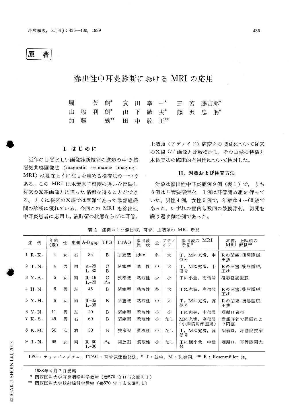
近年の目覚ましい画像診断技術の進歩の中で核磁気共鳴画像法(magnetic resonancc imaging:MRI)は現在とくに注目を集める検査法の一つである。このMRIは水素原子密度の違いを反映し従来のX線画像とは違った情報を得ることができる。とくに従来のX線では困難であった軟部組織間の診断に優れている。今回このMRIを滲出性中耳炎患者に応用し,液貯留の状態ならびに耳管,上咽頭(アデノイド)病変との関係について従来のX線CT画像と比較検討し,その画像の特徴と本検査法の臨床的有用性について検討した。
An applicability of magnetic resonance imaging (MRI) for diagnosing otitis media with effusion (OME) was evaluated. MR images were obtained from 9 patients with OME and were compared with x-ray CT scans.
Effusion was visualized as high-contrast image from adjacent tissues in long SE.
Anatomical and pathological structures of carti-lagenous part of the eustachian tube (ET) were well visualized by MRI while those of osseous part were by CT.
Adenoid tissue was shown as high-contrast image in long SE. Its size and features to sur-rounding tissues were well visualized.
MRI is useful for diagnosing OME in correla-tion between the middle ear and tubo-nasophar-yngeal disorders, especially for defining the pathology of intractable cases.

Copyright © 1989, Igaku-Shoin Ltd. All rights reserved.


