Japanese
English
- 有料閲覧
- Abstract 文献概要
- 1ページ目 Look Inside
はじめに
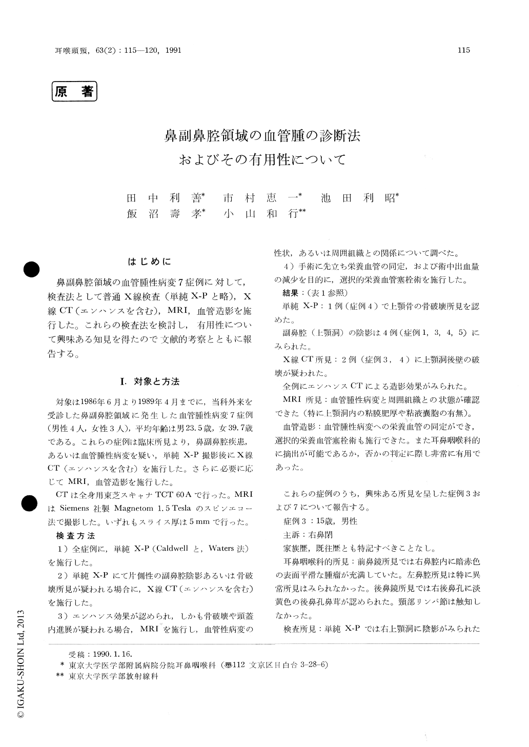
鼻副鼻腔領域の血管腫性病変7症例に対して,検査法として普通X線検査(単純X-Pと略),X線CT (エンハンスを含む),MRI,血管造影を施行した。これらの検査法を検討し,有用性について興味ある知見を得たので文献的考察とともに報告する。
Seven cases of naso-sinal hemangiomatous lesions are reported. Hemangiomas are clinically classified into two types, capillary hemangiomas, and ca-vernous or mixed hemangiomas. CT scan with enhancement reveales abundance of vascularity.MRI well demonstrates interfaces between heman-giomas and normal tissues.
Angiography with selective arterial embolization serves for both identification of feeding artery and decrease in bleeding during surgery.
Judicious use and combination of these diagnostic modalities will lead to more precise evaluation for hemangiomatous lesions in the nose and para-nasal sinuses.
A flow-chart for image diagnosis of these lesions is presented.

Copyright © 1991, Igaku-Shoin Ltd. All rights reserved.


