Japanese
English
- 有料閲覧
- Abstract 文献概要
- 1ページ目 Look Inside
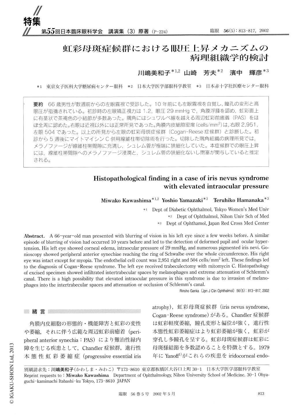
66歳男性が数週前からの左眼霧視で受診した。10年前にも左眼霧視を自覚し,瞳孔の変形と高眼圧が指摘されている。初診時の左眼矯正視力は1.2,眼圧29mmHgで,角膜浮腫を認め,虹彩面上に有茎状で茶褐色の小結節が多数あった。隅角にはシュワルベ線を越える周辺虹彩前癒着(PAS)をほぼ全周に認めた。右眼は近視以外には正常所見であった。角膜内皮細胞密度(cells/mm2)は,右眼2,951,左眼504であった。以上の所見から左眼の虹彩母斑症候群(Cogan-Reese症候群)と診断した。初診から5週後にマイトマイシンC併用線維柱帯切除術を行った。切除した隅角組織の病理所見では,メラノファージが線維柱帯間隙に充満し,シュレム管が極端に狭細化していた。本症候群での眼圧上昇には,線維柱帯間隙へのメラノファージ浸潤と、シュレム管の狭細化ないし閉塞が関与していると推定される。
A 66-year-old man presented with blurring of vision in his left eye since a few weeks before. A similar episode of blurring of vision had occurred 10 years before and led to the detection of deformed pupil and ocular hyper-tension. His left eye showed corneal edema, intraocular pressure of 29mmHg, and numerous pigmented iris nevi. Go-nioscopy showed peripheral anterior synechiae reaching the ring of Schwalbe over the whole circumference. His right eye was intact except for myopia. The endothelial cell count was 2,951 right and 504 cells/mm2 left. These findings led to the diagnosis of Cogan-Reese syndrome. The left eye received trabeculectomy with mitomycin C. Histopathology of excised specimen showed infiltrated intertrabecular spaces by melanophages and extreme attenuation of Schlemm's canal. There is a high possibility that elevated intraocular pressure in this syndrome is due to invasion of melano-phages into the intertrabecular spaces and attenuation or occlusion of Schlemm's canal.

Copyright © 2002, Igaku-Shoin Ltd. All rights reserved.


