Japanese
English
- 有料閲覧
- Abstract 文献概要
- 1ページ目 Look Inside
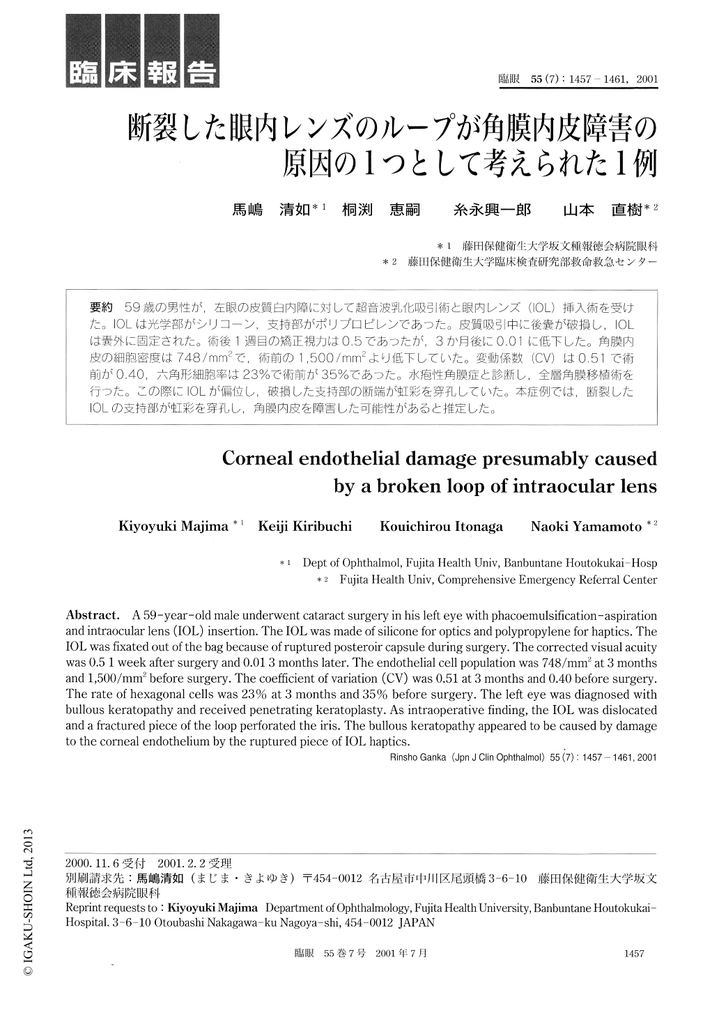
59歳の男性が,左眼の皮質白内障に対して超音波乳化吸引術と眼内レンズ(IOL)挿入術を受けた。IOLは光学部がシリコーン,支持部がポリプロピレンであった。皮質吸引中に後嚢が破損し,IOLは嚢外に固定された。術後1週目の矯正視力は0.5であったが,3か月後に0.01に低下した。角膜内皮の細胞密度は748/mm2で,術前の1,500/mm2より低下していた。変動係数(CV)は0.51で術前が0.40,六角形細胞率は23%で術前が35%であった。水疱性角膜症と診断し,全層角膜移植術を行った。この際にIOLが偏位し,破損した支持部の断端が虹彩を穿孔していた。本症例では,断裂したIOLの支持部が虹彩を穿孔し,角膜内皮を障害した可能性があると推定した。
A 59-year-old male underwent cataract surgery in his left eye with phacoemulsification-aspiration and intraocular lens (IOL) insertion. The IOL was made of silicone for optics and polypropylene for haptics. The IOL was fixated out of the bag because of ruptured posteroir capsule during surgery. The corrected visual acuity was 0.5 1 week after surgery and 0.01 3 months later. The endothelial cell population was 748/mm2 at 3 months and 1,500/mm2 before surgery. The coefficient of variation (CV) was 0.51 at 3 months and 0.40 before surgery. The rate of hexagonal cells was 23% at 3 months and 35% before surgery. The left eye was diagnosed with bullous keratopathy and received penetrating keratoplasty. As intraoperative finding, the IOL was dislocated and a fractured piece of the loop perforated the iris. The bullous keratopathy appeared to be caused by damage to the corneal endothelium by the ruptured piece of IOL haptics.

Copyright © 2001, Igaku-Shoin Ltd. All rights reserved.


