Japanese
English
- 有料閲覧
- Abstract 文献概要
- 1ページ目 Look Inside
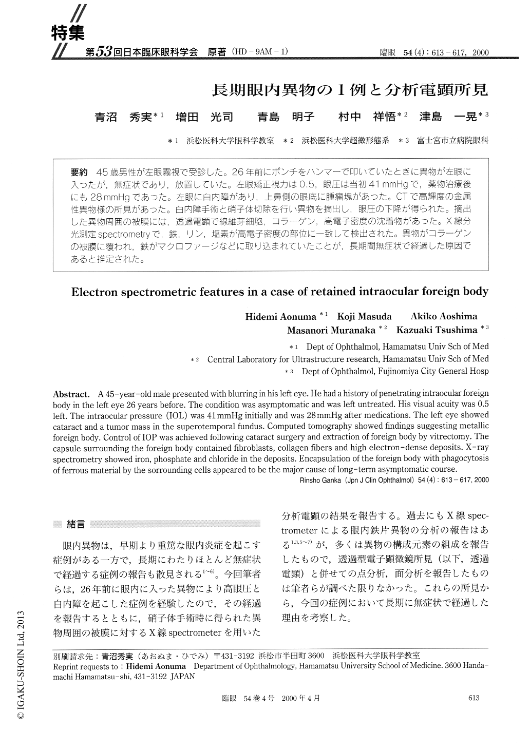
(HD-9AM-1) 45歳男性が左眼霧視で受診した。26年前にポンチをハンマーで叩いていたときに異物が左眼に入ったが,無症状であり,放置していた。左眼矯正視力は0.5,眼圧は当初41mmHgで,薬物治療後にも28mmHgであった。左眼に白内障があり,上鼻側の眼底に腫瘤塊があった。CTで高輝度の金属性異物様の所見があった。白内障手術と硝子体切除を行い異物を摘出し,眼圧の下降が得られた。摘出した異物周囲の被膜には,透過電顕で線維芽細胞,コラーゲン,高電子密度の沈着物があった。X線分光測定spectrometryで,鉄,リン,塩素が高電子密度の部位に一致して検出された。異物がコラーゲンの被膜に覆われ,鉄がマクロファージなどに取り込まれていたことが,長期間無症状で経過した原因であると推定された。
A 45-year-old male presented with blurring in his left eye. He had a history of penetrating intraocular foreign body in the left eye 26 years before. The condition was asymptomatic and was left untreated. His visual acuity was 0.5 left. The intraocular pressure (IOL) was 41 mmHg initially and was 28 mmHg after medications. The left eye showed cataract and a tumor mass in the superotemporal fundus. Computed tomography showed findings suggesting metallic foreign body. Control of TOP was achieved following cataract surgery and extraction of foreign body by vitrectomy. The capsule surrounding the foreign body contained fibroblasts, collagen fibers and high electron-dense deposits. X-ray spectrometry showed iron, phosphate and chloride in the deposits. Encapsulation of the foreign body with phagocytosis of ferrous material by the sorrounding cells appeared to be the major cause of long-term asymptomatic course.

Copyright © 2000, Igaku-Shoin Ltd. All rights reserved.


