Japanese
English
- 有料閲覧
- Abstract 文献概要
- 1ページ目 Look Inside
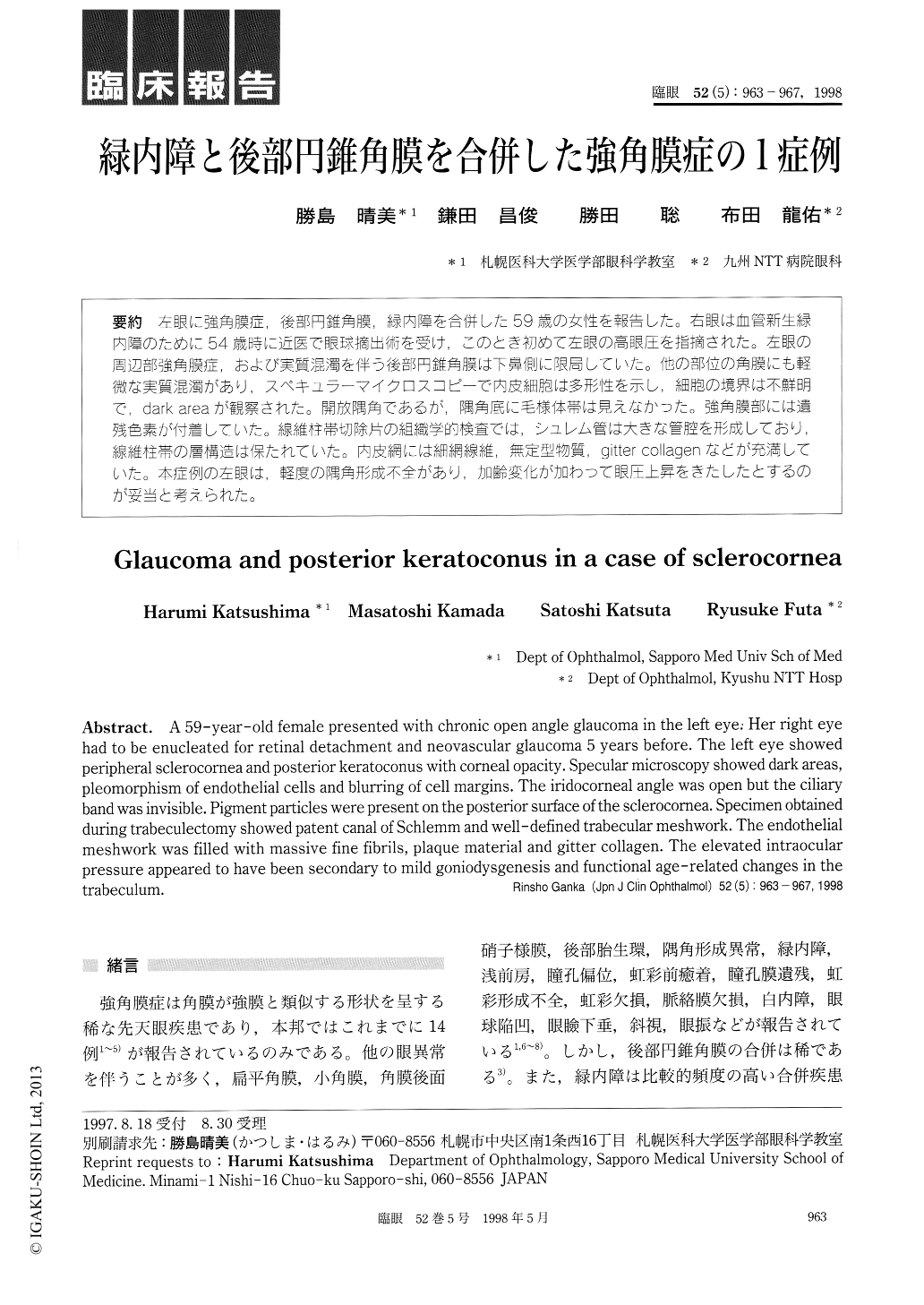
左眼に強角膜症,後部円錐角膜,緑内障を合併した59歳の女性を報告した。右眼は血管新生緑内障のために54歳時に近医で眼球摘出術を受け,このとき初めて左眼の高眼圧を指摘された。左眼の周辺部強角膜症,および実質混濁を伴う後部円錐角膜は下鼻側に限局していた。他の部位の角膜にも軽微な実質混濁があり,スペキュラーマイクロスコピーで内皮細胞は多形性を示し,細胞の境界は不鮮明で,dark areaが観察された。開放隅角であるが,隅角底に毛様体帯は見えなかった。強角膜部には遺残色素が付着していた。線維柱帯切除片の組織学的検査では,シュレム管は大きな管腔を形成しており,線維柱帯の層構造は保たれていた。内皮網には細網線維無定型物質,gitter collagenなどが充満していた。本症例の左眼は,軽度の隅角形成不全があり,加齢変化が加わつて眼圧上昇をきたしたとするのが妥当と考えられた。
A 59-year-old female presented with chronic open angle glaucoma in the left eye: Her right eye had to be enucleated for retinal detachment and neovascular glaucoma 5 years before. The left eye showed peripheral sclerocornea and posterior keratoconus with corneal opacity. Specular microscopy showed dark areas, pleomorphism of endothelial cells and blurring of cell margins. The iridocorneal angle was open but the ciliary band was invisible. Pigment particles were present on the posterior surface of the sclerocornea. Specimen obtained during trabeculectomy showed patent canal of Schlemm and well-defined trabecular meshwork. The endothelial meshwork was filled with massive fine fibrils, plaque material and gitter collagen. The elevated intraocular pressure appeared to have been secondary to mild goniodysgenesis and functional age-related changes in the trabeculum.

Copyright © 1998, Igaku-Shoin Ltd. All rights reserved.


