Japanese
English
- 有料閲覧
- Abstract 文献概要
- 1ページ目 Look Inside
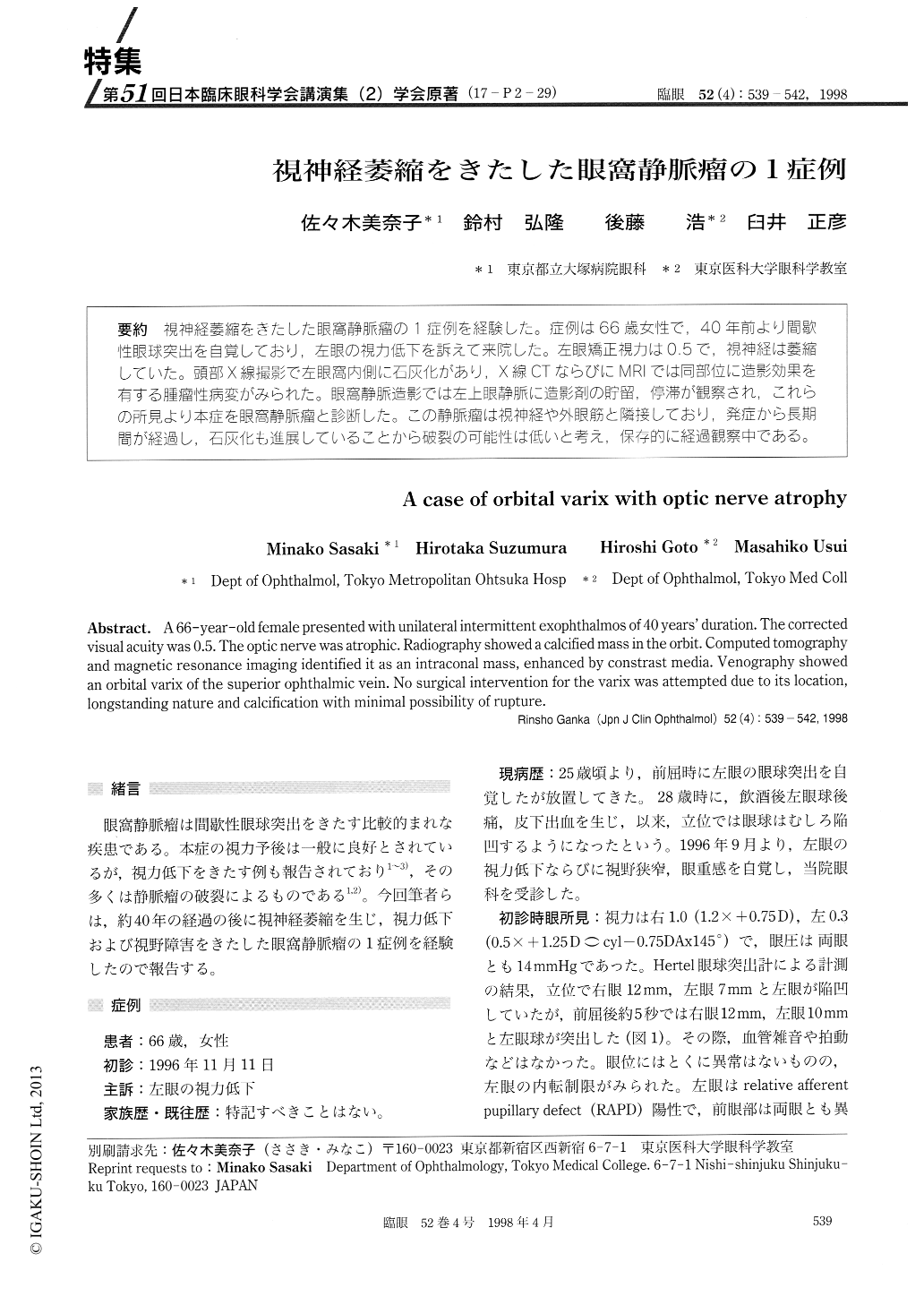
(17-P2-29) 視神経萎縮をきたした眼窩静脈瘤の1症例を経験した。症例は66歳女性で,40年前より間歇性眼球突出を自覚しており,左眼の視力低下を訴えて来院した。左眼矯正視力は0.5で,視神経は萎縮していた。頭部X線撮影で左眼窩内側に石灰化があり,X線CTならびにMRIでは同部位に造影効果を有する腫瘤性病変がみられた。眼窩静脈造影では左上眼静脈に造影剤の貯留,停滞が観察され,これらの所見より本症を眼窩静脈瘤と診断した。この静脈瘤は視神経や外眼筋と隣接しており,発症から長期間が経過し,石灰化も進展していることから破裂の可能性は低いと考え,保存的に経過観察中である。
A 66-year-old female presented with unilateral intermittent exophthalmos of 40 years' duration. The corrected visual acuity was 0.5. The optic nerve was atrophic. Radiography showed a calcified mass in the orbit. Computed tomography and magnetic resonance imaging identified it as an intraconal mass, enhanced by constrast media. Venography showed an orbital varix of the superior ophthalmic vein. No surgical intervention for the varix was attempted due to its location, longstanding nature and calcification with minimal possibility of rupture.

Copyright © 1998, Igaku-Shoin Ltd. All rights reserved.


