Japanese
English
- 有料閲覧
PPV購入案内
1,320円 (1,200円+税10%) こちらは、217ページから220ページの文献です。購入には医書.jp本会員登録が必要です。
会員登録完了後に改めて上記「購入サイトへ」を選択してください。
または「購入サイトへ」選択後に「購入ログイン」、「新規会員登録」の順に進んでください。
会員登録について詳しくはをご確認ください。
- Abstract 文献概要
- 1ページ目 Look Inside
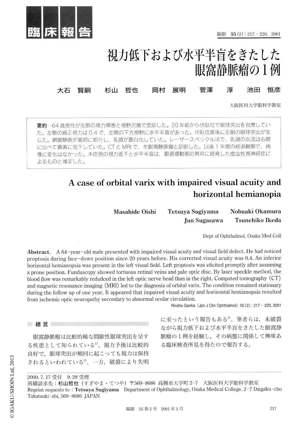
64歳男性が左眼の視力障害と視野欠損で受診した。20年前から伏臥位で眼球突出を自覚していた。左眼の矯正視力は0.4で,左眼の下方視野に水平半盲があった。伏臥位直後に左眼の眼球突出が生じた。網膜静脈が著明に蛇行し,乳頭が蒼白化していた。レーザースペックル法で,乳頭の血流は右眼に比べて顕著に低下していた。CTとMRIで,左眼窩静脈瘤と診断した。以後1年間の経過観察で,病像に変化はなかった。本症例の視力低下と水平半盲は,眼循環動態の異常に続発した虚血性視神経症によるものと推定した。
A 64-year-old male presented with impaired visual acuity and visual field defect. He had noticed proptosis during face-down position since 20 years before. His correrted visual acuity was 0.4. An inferior horizontal hemianopsia was present in the left visual field. Left proptosis was elicited promptly after assuming a prone position. Funduscopy showed tortuous retinal veins and pale optic disc. By laser speckle method, the blood flow was remarkably redudced in the left optic nerve head than in the right. Computed tomography (CT) and magnetic resonance imaging (MRI) led to the diagnosis of orbital varix. The condition remained stationary during the follow up of one year. It appeared that impaired visual acuity and horizontal hemianopsia resulted from ischemic optic neuropathy secondary to abnormal ocular circulation.
Copyright © 2001, Igaku-Shoin Ltd. All rights reserved.