Japanese
English
- 有料閲覧
- Abstract 文献概要
- 1ページ目 Look Inside
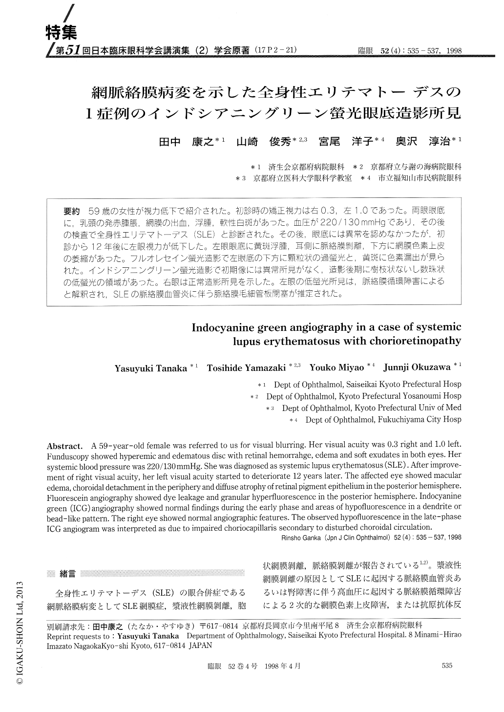
(17P2-21) 59歳の女性が視力低下で紹介された。初診時の矯正視力は右0.3,左1.0であった。両眼眼底に,乳頭の発赤腫脹,網膜の出血,浮腫,軟性白斑があった。血圧が220/130 mmHgであり,その後の検査で全身性エリテマトーデス(SLE)と診断された。その後,眼底には異常を認めなかったが,初診から12年後に左眼視力が低下した。左眼眼底に黄斑浮腫,耳側に脈絡膜剥離,下方に網膜色素上皮の萎縮があった。フルオレセイン螢光造影で左眼底の下方に顆粒状の過螢光と,黄斑に色素漏出が見られた。インドシアニングリーン螢光造影で初期像には異常所見がなく,造影後期に樹枝状ないし数珠状の低螢光の領域があった。右眼は正常造影所見を示した。左眼の低螢光所見は,脈絡膜循環障害によると解釈され,SLEの脈絡膜血管炎に伴う脈絡膜毛細管板閉塞が推定された。
A 59-year-old female was referred to us for visual blurring. Her visual acuity was 0.3 right and 1.0 left. Funduscopy showed hyperemic and edematous disc with retinal hemorrahge, edema and soft exudates in both eyes. Her systemic blood pressure was 220/130 mmHg. She was diagnosed as systemic lupus erythematosus (SLE). After improve-ment of right visual acuity, her left visual acuity started to deteriorate 12 years later. The affected eye showed macular edema, choroidal detachment in the periphery and diffuse atrophy of retinal pigment epithelium in the posterior hemisphere. Fluorescein angiography showed dye leakage and granular hyperfluorescence in the posterior hemisphere. Indocyanine green (ICG) angiography showed normal findings during the early phase and areas of hypofluorescence in a dendrite or bead-like pattern. The right eye showed normal angiographic features. The observed hypofluorescence in the late-phase ICG angiogram was interpreted as due to impaired choriocapillaris secondary to disturbed choroidal circulation.
Copyright © 1998, Igaku-Shoin Ltd. All rights reserved.