Japanese
English
- 有料閲覧
- Abstract 文献概要
- 1ページ目 Look Inside
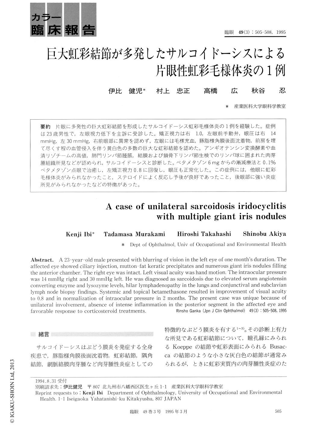
片眼に多発性の巨大虹彩結節を形成したサルコイドーシス虹彩毛様体炎の1例を経験した。症例は23歳男性で,左眼視力低下を主訴に受診した。矯正視力は右 1.0,左眼前手動弁,眼圧は右 14mmHg,左30 mmHg,右前眼部に異常を認めず,左眼には毛様充血,豚脂様角膜後面沈着物,前房を埋て尽くす程の血管侵入を伴う黄白色の多数の巨大な虹彩結節を認めた。アンギオテンシン変換酵素や血清リゾチームの高値,肺門リンパ節腫脹,結膜および鎖骨下リンパ節生検でのリンパ球に囲まれた肉芽腫組織所見などが認められ,サルコイドーシスと診断した。ベタメタゾン6mgからの漸減療法と0.1%ベタメタゾン点眼で治癒し,左矯正視力0.8に回復し,眼圧も正常化した。この症例には,他眼に虹彩毛様体炎がみられなかったこと,ステロイドによく反応し予後が良好であったこと,後眼部に強い炎症所見がみられなかったなどの特徴があった。
A 23-year-old male presented with blurring of vision in the left eye of one month's duration. The affected eye showed ciliary injection, mutton-fat keratic precipitates and numerous giant iris nodules filling the anterior chamber. The right eye was intact. Left visual acuity was hand motion. The intraocular pressure was 14 mmHg right and 30 mmHg left. He was diagnosed as sarcoidosis due to elevated serum angiotensin converting enzyme and lysozyme levels, hilar lymphadenopathy in the lungs and conjunctival and subclavian lymph node biopsy findings. Systemic and topical betamethasone resulted in improvement of visual acuity to 0.8 and in normalization of intraocular pressure in 2 months. The present case was unique because of unilateral involvement, absence of intense inflammation in the posterior segment in the affected eye and favorable response to corticosteroid treatments.

Copyright © 1995, Igaku-Shoin Ltd. All rights reserved.


