Japanese
English
- 有料閲覧
- Abstract 文献概要
- 1ページ目 Look Inside
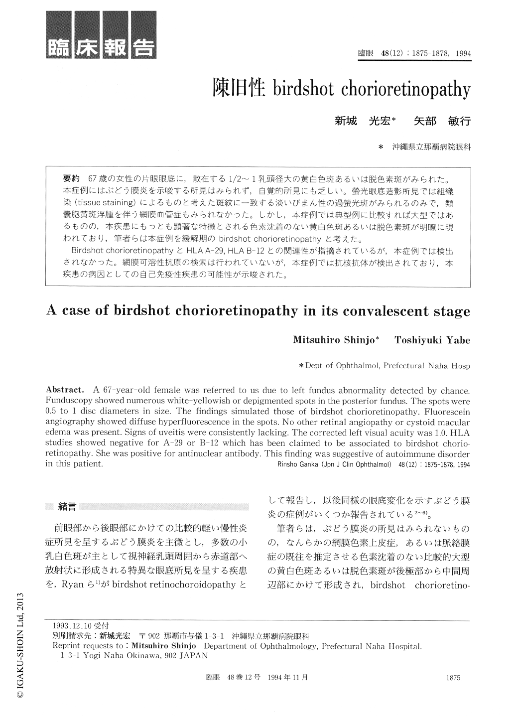
67歳の女性の片眼眼底に,散在する1/2〜1乳頭径大の黄白色斑あるいは脱色素斑がみられた。本症例にはぶどう膜炎を示唆する所見はみられず,自覚的所見にも乏しい。螢光眼底造影所見では組織染(tissue staining)によるものと考えた斑紋に一致する淡いびまん性の過螢光斑がみられるのみで,類嚢胞黄斑浮腫を伴う網膜血管症もみられなかった。しかし,本症例では典型例に比較すれば大型ではあるものの,本疾患にもっとも顕著な特徴とされる色素沈着のない黄白色斑あるいは脱色素斑が明瞭に現われており,筆者らは本症例を緩解期のbirdshot chorioretinopathyと考えた。
Birdshot chorioretinopathyとHLA A−29,HLA B−12との関連性が指摘されているが,本症例では検出されなかった。網膜可溶性抗原の検索は行われていないが,本症例では抗核抗体が検出されており,本疾患の病因としての自己免疫性疾患の可能性が示唆された。
A 67-year-old female was referred to us due to left fundus abnormality detected by chance. Funduscopy showed numerous white-yellowish or depigmented spots in the posterior fundus. The spots were 0.5 to 1 disc diameters in size. The findings simulated those of birdshot chorioretinopathy. Fluorescein angiography showed diffuse hyperfluorescence in the spots. No other retinal angiopathy or cystoid macular edema was present. Signs of uveitis were consistently lacking. The corrected left visual acuity was 1.0. HLA studies showed negative for A-29 or B-12 which has been claimed to be associated to birdshot chorio-retinopathy. She was positive for antinuclear antibody. This finding was suggestive of autoimmune disorder in this patient.

Copyright © 1994, Igaku-Shoin Ltd. All rights reserved.


