Japanese
English
- 有料閲覧
- Abstract 文献概要
- 1ページ目 Look Inside
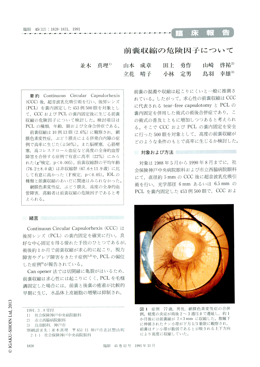
Continuous Circular Capsulorhexis(CCC)後,超音波乳化吸引術を行い,後房レンズ(PCL)を嚢内固定した453例500眼を対象として,CCCおよびPCLの嚢内固定後に生じる前嚢収縮の危険因子について検討した。検討項目はPCLの種類,年齢,眼および全身合併症である。
前嚢収縮は10例13眼(2.6%)に観察され,網膜色素変性症,ぶどう膜炎による併発白内障の症例で高率に生じた(≧50%)。また脳梗塞,心筋梗塞,高コレステロール血症など高度の全身的血管障害を合併する症例で有意に高率(22%)にみられた(χ2検定,p<0.005)。前嚢収縮群の平均年齢(76.2±8.0歳)は非収縮群(67.6±11.9歳)に比して有意に高かった(F検定,p<0.05)。IOLの種類と前嚢収縮のあいだに関連はみられなかった。
網膜色素変性症,ぶどう膜炎,高度の全身的血管障害,高齢者は前嚢収縮の危険因子であると考えられる。
We evaluated the risk factors for anterior capsu-lar shrinkage in 500 eyes after intraocular lens implantation. All the eyes underwent phacoemul-sification/aspiration after continuous circular cap-sulorhexis of about 5mm in diameter. We used posterior chamber lens of 6 or 6.5mm and em-ployed in-the-bag fixation technique. Anterior cap-sular shrinkage was judged as present when the anterior capsular edge was visible in the pupillaryarea by slitlamp examination without myadriatics.
Anterior capsular shrinkage was present in 13 eyes, 2.6%. There was a high incidence of shrinkage in eyes with uveitis or pigmentary retinal dystro-phy. Severe systemic vascular disorders, including myocardial infarction, brain infarction and hyper-cholesteremia, were another contribution factor for shrinkage. The age of cases who developed shrink-age averaged 76.2±8.0 years, which was significant-ly higher than 67.9±11.9 years for those who failed to develop shrinkage. The size and type of intraocu-lar lens did not affect the incidence of shrinkage.

Copyright © 1991, Igaku-Shoin Ltd. All rights reserved.


