Japanese
English
- 有料閲覧
- Abstract 文献概要
- 1ページ目 Look Inside
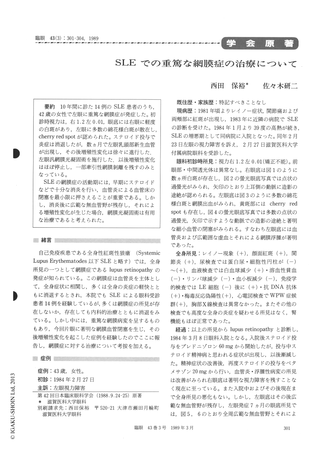
10年間に診た14例のSLE患者のうち,42歳の女性で左眼に重篤な網膜症が発症した。初診時視力は,右1.2左0.01,眼底には右眼に軽度の白斑があり,左眼に多数の綿花様白斑が散在し,cherry red spotが認められた。ステロイド投与で炎症は消退したが,数ヵ月で左眼乳頭部新生血管が出現し,その後増殖性変化は徐々に進行した.左眼汎網膜光凝固術を施行した.以後増殖性変化はほぼ停止し,一部牽引性網膜剥離を残すのみとなっている。
SLEの網膜症の活動期には,早期にステロイドなどで十分な消炎を行い,血管炎による血管床の閉塞を最小限に押さえることが重要である。しかし,消炎後に広範な無血管野が残存し,それによる増殖性変化が生じた場合,網膜光凝固術は有用な治療であると考えられた。
A 42-year-old woman presented with advanced retinopathy in both eyes. She had a history of systemic lupus erythematosus of 3 years' duration. The visual acuity was 1.2 right and 0.01 left. Numerous exudates were seen in the right fundus. Numerous cotton-wool patches and macular cherry red spot were seen in the left fundus.
Systemic corticosteroid treatment was started to control the inflammation. After 7 months, neovas-cularization started to develop from the optic disc in the left eye. Panretinal photocoagulation was performed to treat the avascular retinal area. Marked reduction in proliferative changes resulted after photocoagulation.
We propose that systemic corticosteroid is in-dicated to suppress angiitis and to prevent further obliteration of retinal vessels. If proliferative changes appear secondary to the presence of exten-sive avascular retinal areas, photocoagulation is the treatment of choice to induce regression of neovascularization.

Copyright © 1989, Igaku-Shoin Ltd. All rights reserved.


