Japanese
English
- 有料閲覧
- Abstract 文献概要
- 1ページ目 Look Inside
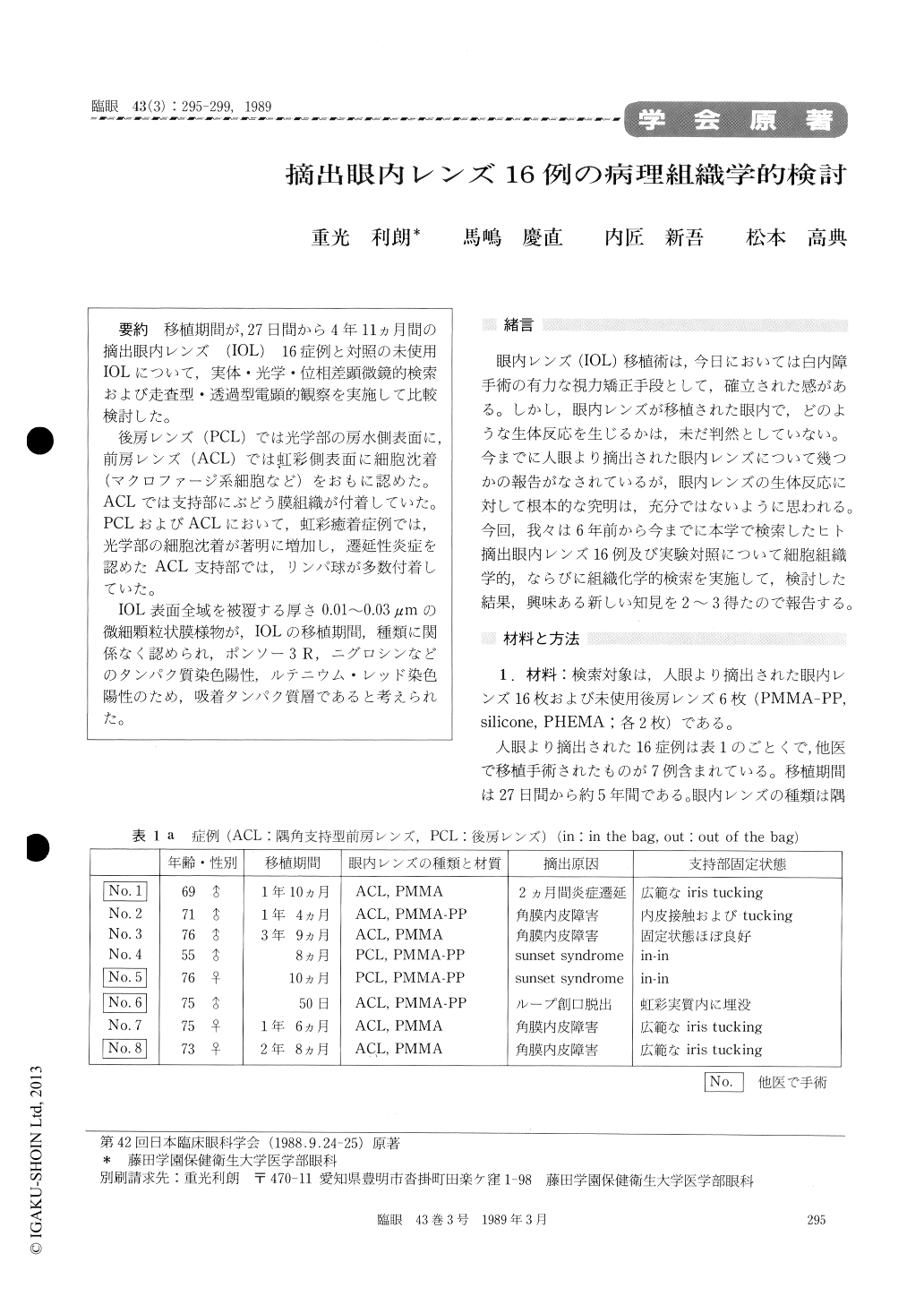
移植期間が,27日間から4年11ヵ月間の摘出眼内レンズ (IOL)16症例と対照の未使用IOLについて,実体・光学・位相差顕微鏡的検索および走査型・透過型電顕的観察を実施して比較検討した。
後房レンズ(PCL)では光学部の房水側表面に,前房レンズ(ACL)では虹彩側表面に細胞沈着(マクロファージ系細胞など)をおもに認めた。ACLでは支持部にぶどう膜組織が付着していた。PCLおよびACLにおいて,虹彩癒着症例では,光学部の細胞沈着が著明に増加し,遷延性炎症を認めたACL支持部では,リンパ球が多数付着していた。
IOL表面全域を被覆する厚さ0.01〜0.03μmの微細顆粒状膜様物が,IOLの移植期間,種類に関係なく認められ,ポンソー3R,ニグロシンなどのタンパク質染色陽性,ルテニウム・レッド染色陽性のため,吸着タンパク質層であると考えられた。
We evaluated the surface structure of 16 surgi-cally removed intraocular lenses (IOLs). The IOLs had stayed in the eye for periods ranging from 27 days to 5 years. We employed stereomicroscope, light and phase-contrast microscopes and scanning and transmission electron microscopes.
Cell deposits, mainly macrophages, were present on the anterior face of posterior chamber lenses (PCLs) and on the posterior face of anterior cham-ber lenses (ACLs). Uveal tissue was adherent to the haptics of ACLs. Cell deposits in the optic portion were more pronounced in cases of iris adhesion to the IOLs. Numerous lymphocytes adhered to the haptics of ACLs in eyes with protracted intraocular inflammation.
An ultrathin granular membrane, 10 to 30 nm in thickness, covered the both surfaces of IOLs regard-less of the type and duration of IOLs. This mem-brane-like structure showed histochemical charac-teristics suggestive of adsorbed proteinous layer.

Copyright © 1989, Igaku-Shoin Ltd. All rights reserved.


