Japanese
English
- 有料閲覧
- Abstract 文献概要
- 1ページ目 Look Inside
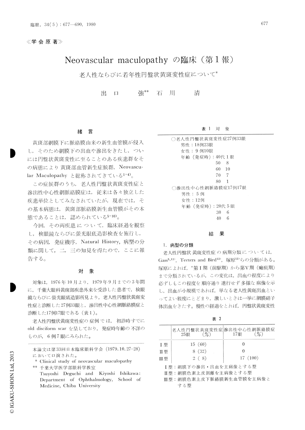
(1) Neovascular maculopathy (老人性円盤状黄斑変性症ならびに滲出性中心性網脈絡膜症)の活動期病変は,検眼鏡所見ならびに螢光眼底造影所見より,次の3型に大別された。
Ⅰ型:網膜下の滲出・出血を主病像とする型
Ⅱ型:網膜色素上皮剥離を主病像とする型
Ⅲ型:網膜色素上皮下脈絡膜新生血管膜を主病像とする型
(2)長期間の臨床経過を観察できた滲出性中心性網脈絡膜症では,60%は脈絡膜新生血管膜の退縮・瘢痕化に至り,軽度の瘢痕と周囲の色素上皮の萎縮を残したが,良好な視力を回復した。40%は新生血管膜の増大と,それによる滲出が遷延化し,若年性円盤状黄斑変性症とでもいえる病像を呈し,ついには,より大きな網脈絡膜変性巣を残し,高度の視力障害を残した。
(3)その他の所見として,老人性円盤状黄斑変性症では,黄斑部ドルーゼ様変化または網膜色素上皮の変性萎縮像を呈していた例は,患眼では50%,fellow eyeでは52%にみられた。
滲出性中心性網脈絡膜症の患眼においては,小円形網脈絡膜萎縮巣を伴っていた例は35.3%にみられ,近視眼が88.2%の高率に含まれていた。
We studied 33 eyes of 27 patients with senile disciform macular degeneration and 17 eyes of 17 patients with central exudative chorioretinopathy by means of funduscopy and serial fluoresein angiography.
The results were as follows:
1) The funduscopic pictures of neovasular maculo-pathy were classified into 3 groups.
Ⅰ. Type with subretinal exudate and hemor-rhage
Ⅱ. Type with pigment epithelial detachment
Ⅲ. Type with subpigment epithelial neova-scular membrane

Copyright © 1980, Igaku-Shoin Ltd. All rights reserved.


