Japanese
English
- 有料閲覧
- Abstract 文献概要
- 1ページ目 Look Inside
緒 言
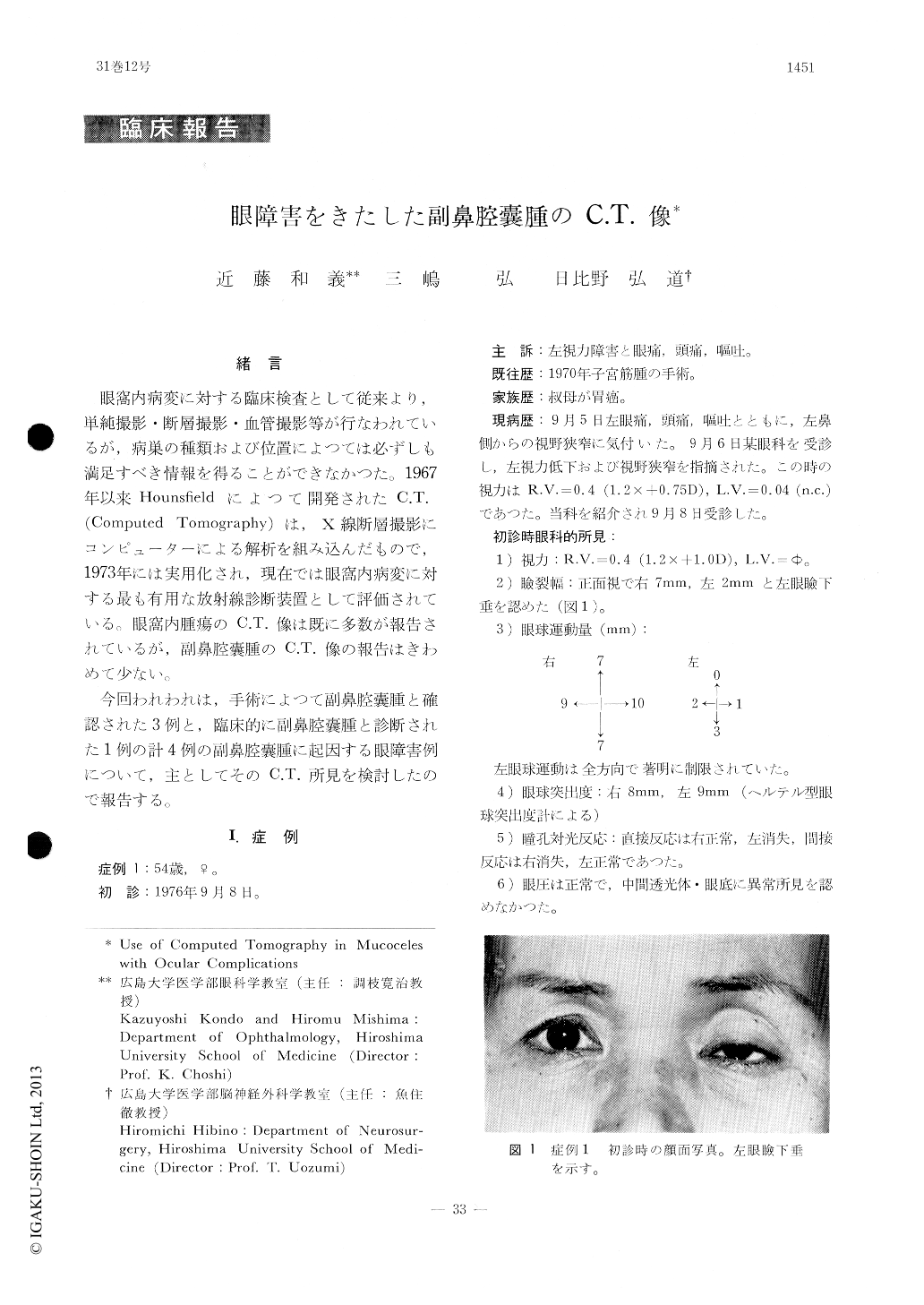
眼窩内病変に対する臨床検査として従来より,単純撮影。断層撮影・血管撮影等が行なわれているが,病巣の種類および位置によつては必ずしも満足すべき情報を得ることができなかつた。1967年以来Hounsfieldによつて開発されたC.T.(Computed Tomography)は,X線断層撮影にコンピューターによる解析を組み込んだもので,1973年には実用化され,現在では眼窩内病変に対する最も有用な放射線診断装置として評価されている。眼窩内腫瘍のC.T.像は既に多数が報告されているが,副鼻腔嚢腫のC.T.像の報告はきわめて少ない。
今回われわれは,手術によつて副鼻腔嚢腫と確認された3例と,臨床的に副鼻腔嚢腫と診断された1例の計4例の副鼻腔嚢腫に起因する眼障害例について,主としてそのC.T.所見を検討したので報告する。
We examined 4 cases of ocular complicationsdue to mucoceles of the paranasal sinus by means of computed tomography (EMI-1000). Ocular manifestations of one case consisted of unilateral blindness, ophthalmoplegia, headache and bleph-aroptosis, so that we diagosed this case as orbital apex syndrome initially. Epiphora was the sole ocular manifestation in another case. Unilateral proptosis was the chief clinical feature in the other two cases. In all the four cases, computed tomography readily led to the detection of the presence of homogeneous space-occupying lesions in the paranasal sinus and the orbit.

Copyright © 1977, Igaku-Shoin Ltd. All rights reserved.


