Japanese
English
- 有料閲覧
- Abstract 文献概要
- 1ページ目 Look Inside
緒 言
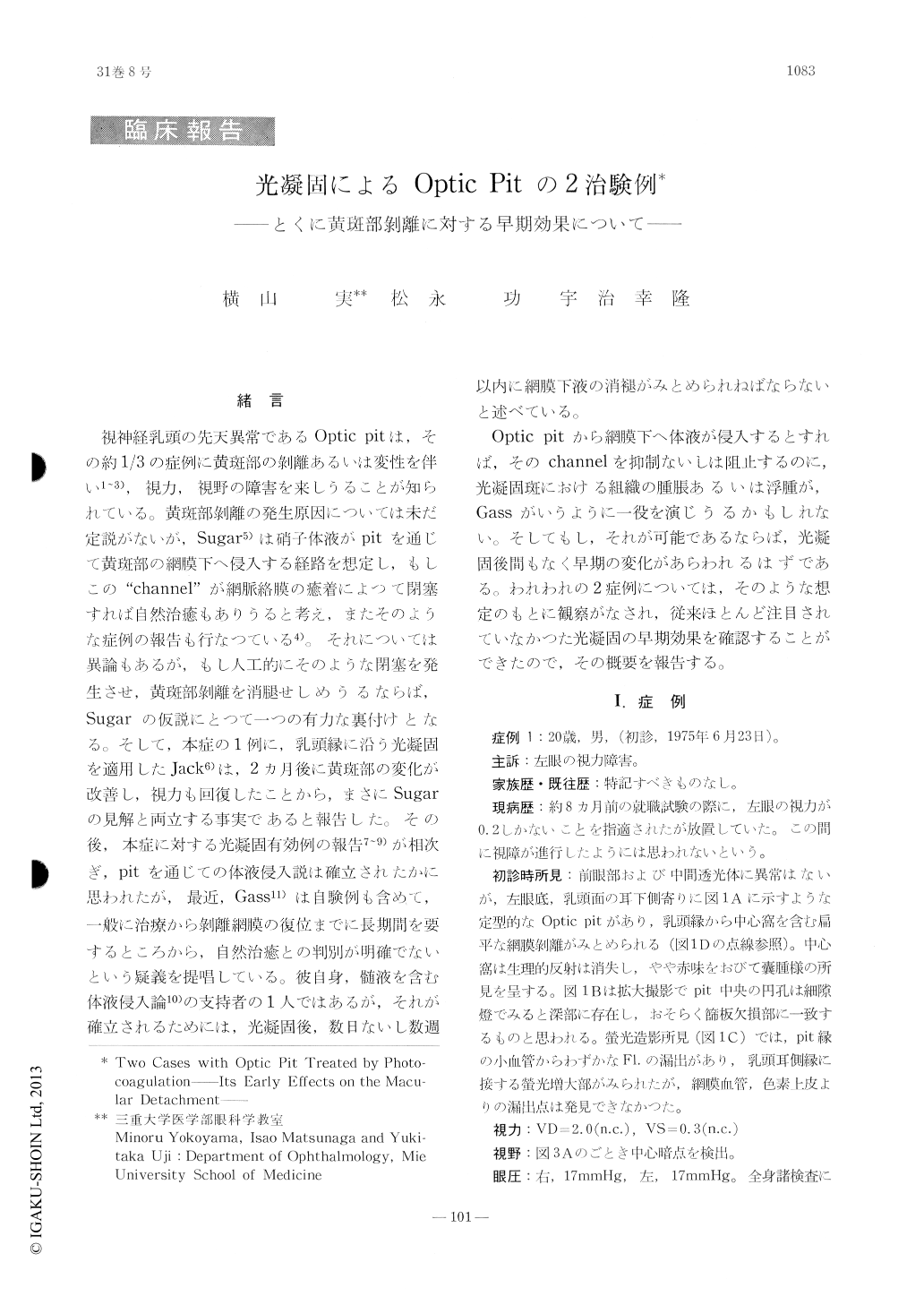
視神経乳頭の先天異常であるOptic pitは,その約1/3の症例に黄斑部の剥離あるいは変性を伴い1〜3),視力,視野の障害を来しうることが知られている。黄斑部剥離の発生原因については未だ定説がないが,Sugar5)は硝子体液がpitを通じて黄斑部の網膜下へ侵入する経路を想定し,もしこの"channel"が網脈絡膜の癒着によつて閉塞すれば自然治癒もありうると考え,またそのような症例の報告も行なつている4)。それについては異論もあるが,もし人工的にそのような閉塞を発生させ,黄斑部剥離を消腿せしめうるならば,Sugarの仮説にとつて一つの有力な裏付けとなる。そして,本症の1例に,乳頭縁に沿う光凝固を適用したJack6)は,2カ月後に黄斑部の変化が改善し,視力も回復したことから,まさにSugarの見解と両立する事実であると報告した。その後,本症に対する光凝固有効例の報告7〜9)が相次ぎ,pitを通じての体液侵入説は確立されたかに思われたが,最近,Gass11)は自験例も含めて,一般に治療から剥離網膜の復位までに長期間を要するところから,自然治癒との判別が明確でないという疑義を提唱している。彼自身,髄液を含む体液侵入論10)の支持者の1人ではあるが,それが確立されるためには,光凝固後,数日ないし数週以内に網膜下液の消褪がみとめられねばならないと述べている。
Congenital pit of the optic nervehead associ-ated with serous detachment of posterior retina was seen in two males aged 20 and 7 years each. Patients were treated by xenon photoco-agulation along the disc margin adjacent to the retinal detachment. Almost complete reattach-ment of the retina resulted in the first case, while the reattachment was incomplete in the second case leaving a localized detached area around the optic disc.
The observed effects of photocoagulation in these two cases became manifest in two, early and late, phases.

Copyright © 1977, Igaku-Shoin Ltd. All rights reserved.


