Japanese
English
- 有料閲覧
- Abstract 文献概要
- 1ページ目 Look Inside
緒 言
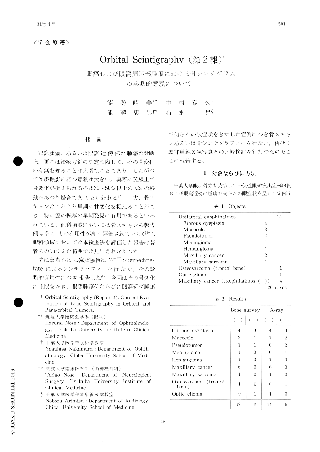
眼窩腫瘍,あるいは眼窩近傍部の腫瘍の診断上,更には治療方針の決定に際して,その骨変化の有無を知ることは大切なことであり,したがつてX線撮影の持つ意義は大きい。実際にX線上で骨変化が捉えられるのは30〜50%以上のCaの移動があつた場合であるといわれる1)。一方,骨スキャンはこれより早期に骨変化を捉えることができ,特に癌の転移の早期発見に有用であるといわれている。他科領域においては骨スキャンの報告例も多く,その有用性が高く評価されているが2〜5)眼科領域においては本検査法を評価した報告は著者らの知りえた範囲では見出されなかつた。
先に著者らは眼窩腫瘍例に99mTc-pertechne-tateによるシンチグラフィーを行ない,その診断的有用性につき報告した6)。今回はその骨変化に主眼をおき中眼窩腫瘍例ならびに眼窩近傍腫瘍で何らかの眼症状をきたした症例につき骨スキャンあるいは骨シンチグラフィーを行ない,併せて頭部単純X線写真との比較検討を行なつたのでここに報告する。
This paper reports the results of clinicalstudies of bone scintigrams with Tc-diphosph-onate and comparisons of the scintigrams with the skull X-ray films in those cases. The pati-ents studied in this series were sixteen cases of unilateral exophthalmos by the tumors ori-ginating from orbit or para-nasal sinuses and four cases of para-orbital tumors.
Scintigraphic findings were abnormal in seven-teen cases. Skull X-ray findings were abnormal in fourteen cases, and they all but one also showed abnormal scintigrams. The detected bone changes were wider and more distinct in the latter than in the former.

Copyright © 1977, Igaku-Shoin Ltd. All rights reserved.


