Japanese
English
臨床実験
広角眼底カメラによる螢光撮影について
Fluorescein Angiography with a Wide Angle Fundus Camera
田中 宏和
1
,
野寄 達司
1
,
馬場 賢一
1
,
藤田 邦彦
1
,
加藤 尚臣
2
,
田尾 森郎
2
Hirokazu Tanaka
1
,
Satoshi Noyori
1
,
Ken-ichi Baba
1
,
Kunihiko Fujita
1
,
Naomi Kato
2
,
Moriro Tawo
2
1順天堂大学医学部眼科学教室
2日本光学株式会社
1Department of Ophthalmology, Juntendo Univcrsity School of Medicine
2Nippon Kogaku Co.
pp.213-215
発行日 1972年2月15日
Published Date 1972/2/15
DOI https://doi.org/10.11477/mf.1410204730
- 有料閲覧
- Abstract 文献概要
- 1ページ目 Look Inside
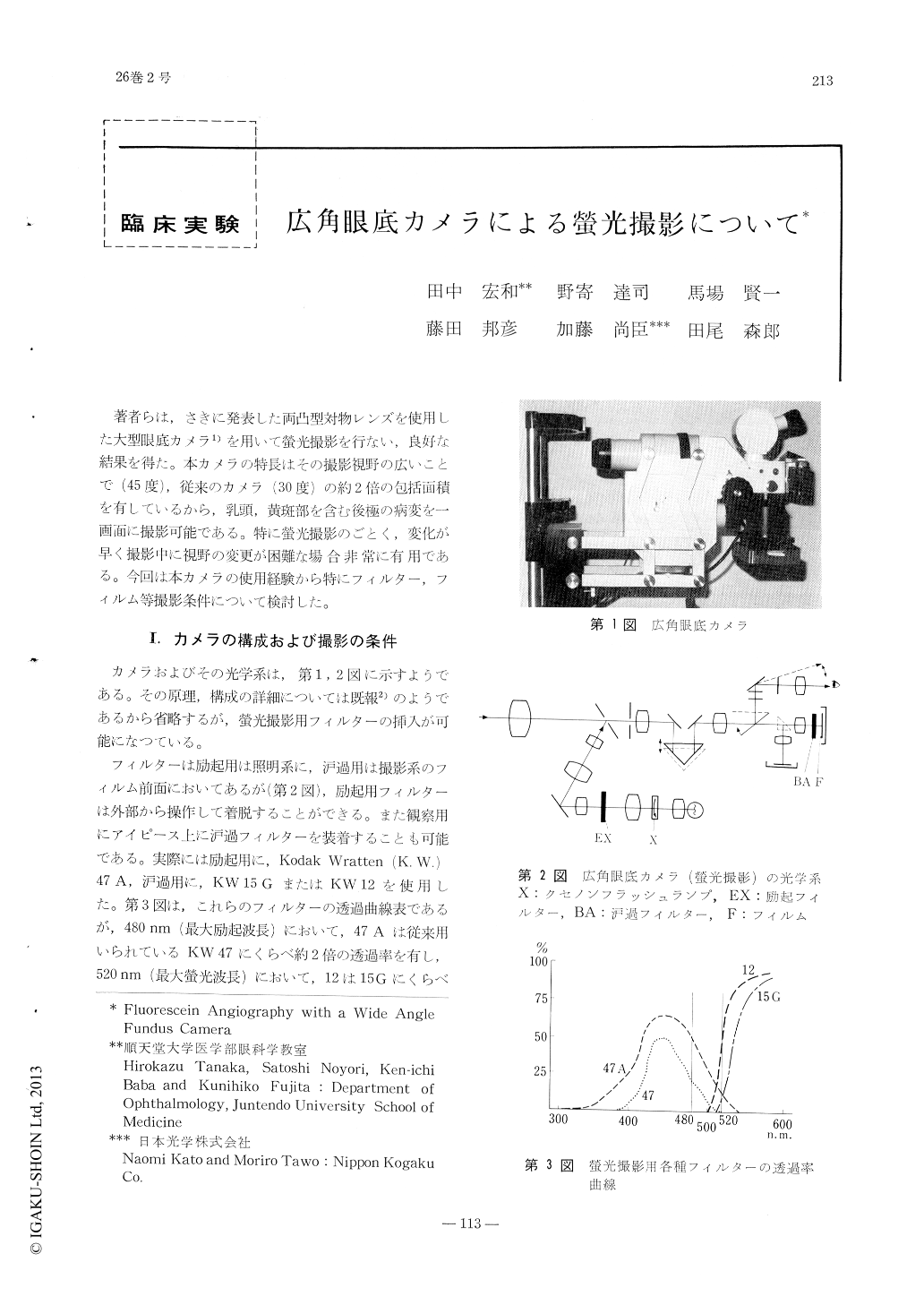
著者らは,さきに発表した両凸型対物レンズを使用した大型眼底カメラ1)を用いて螢光撮影を行ない,良好な結果を得た。本カメラの特長はその撮影視野の広いことで(45度),従来のカメラ(30度)の約2倍の包括面積を有しているから,乳頭,黄斑部を含む後極の病変を一画面に撮影可能である。特に螢光撮影のごとく,変化が早く撮影中に視野の変更が困難な場合非常に有用である。今回は本カメラの使用経験から特にフィルター,フイルム等撮影条件について検討した。
Fluorescein angiography with widefield fundus camera (45°) is discussed. The camera can cover a fundus area twice as wide as the ordinary fundus cameras. The whole posterior fundus including the optic disc and the macula can be documented in one picture.
The method promises to be of value in exa-mining widespread fundus lesions by means of fluorescein angiography.

Copyright © 1972, Igaku-Shoin Ltd. All rights reserved.


