Japanese
English
- 有料閲覧
- Abstract 文献概要
- 1ページ目 Look Inside
緒言
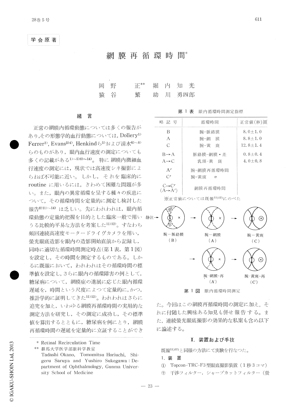
正常の網膜内循環動態については多くの報告があり,その形態学的血行動態については,Dollery1)Ferrer2), Evans3)4), Henkindら5)および清水6)〜8)らのものがあり,眼内血行速度の測定についても多くの記載がある1)〜5)8)〜14)。特に網膜内微細血行速度の測定には,現状では高速度シネ撮影によらねば不可能に近い。しかし,それを臨床的にroutineに用いるには,きわめて困難な問題が多い。また,眼内の異常循環を呈する種々の疾患について,その循環時間を定量的に測定し検討した報告8)11)〜14)は乏しい。先にわれわれは,眼内循環動態の定量的把握を目的とした臨床一般で用いうる比較的平易な方法を考案した11)12)。すなわち頻回連続高速度モータードライヴカメラを用い,螢光眼底造影を眼内の造影開始直前から記録し,同時に適切な循環時間測定時点(第1表,第1図)を設定し,その時間を測定するものである。しかるに既報において,われわれはその循環時間の標準値を設定し,さらに眼内の循環障害の例として,糖尿病について,網膜症の進展に応じた眼内循環遅延を,時間という尺度によつて定量的に,かつ,推計学的に証明してきた11)12)。
The retinal recirculation time was measured in normal and diabetic subjects by means of rapid serial fluorescein angiography for prolon-ged periods. Automatic rapid dye injector and self-winding, long-strip camera with the ca-pacity of photographing 250 successive frames were used as special technical features.
The retinal recirculation time is defined as the time interval between the initial and thesecond appearance of the dye bolus in the cen-tral retinal artery at the optic nervehead. The value was 10.9±1.5 sec in normal 9 subjects and coincided well with the arm-to-retina circu-lation time (8.9±1.1sec).

Copyright © 1974, Igaku-Shoin Ltd. All rights reserved.


