Japanese
English
- 有料閲覧
PPV購入案内
1,320円 (1,200円+税10%) こちらは、1535ページから1542ページの文献です。購入には医書.jp本会員登録が必要です。
会員登録完了後に改めて上記「購入サイトへ」を選択してください。
または「購入サイトへ」選択後に「購入ログイン」、「新規会員登録」の順に進んでください。
会員登録について詳しくはをご確認ください。
- Abstract 文献概要
- 1ページ目 Look Inside
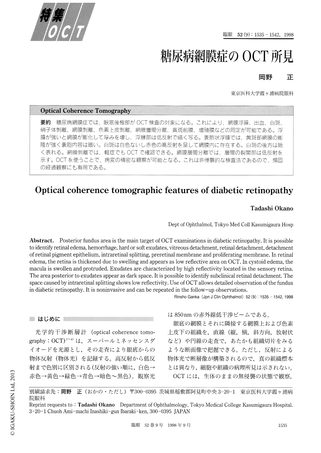
糖尿病網膜症では,眼底後極部がOCT検査の対象になる。これにより,網膜浮腫,出血,白斑,硝子体剥離,網膜剥離,色素上皮剥離,網膜層間分離,黄斑前膜,増殖膜などの同定が可能である。浮腫が強いと網膜が膨化して厚みを増し,浮腫部は低反射で暗く写る。嚢胞状浮腫では,黄斑部網膜の膨隆が強く嚢胞内容は暗い。白斑は白色ないし赤色の高反射を呈して網膜内に存在する。白斑の後方は暗く表れる。網膜剥離では,軽症でもOCTで確認できる。網膜層間分離では,層間の裂開部は低反射を示す。OCTを使うことで,病変の精密な観察が可能となる。これは非侵襲的な検査法であるので,頻回の経過観察にも有用である。
Posterior fundus area is the main target of OCT examinations in diabetic retinopathy. It is possible to identify retinal edema, hemorrhage, hard or soft exudates, vitreous detachment, retinal detachment, detachment of retinal pigment epithelium, intraretinal splitting, preretinal membrane and proliferating membrane. In retinal edema, the retina is thickened due to swelling and appears as low reflective area on OCT. In cystoid edema, the macula is swollen and protruded. Exudates are characterized by high reflectivity located in the sensory retina. The area posterior to exudates appear as dark space. It is possible to identify subclinical retinal detachment. The space caused by intraretinal splitting shows low reflectivity. Use of OCT allows detailed observation of the fundus in diabetic retinopathy. It is noninvasive and can be repeated in the follow-up observations.
Copyright © 1998, Igaku-Shoin Ltd. All rights reserved.