Japanese
English
- 有料閲覧
PPV購入案内
1,320円 (1,200円+税10%) こちらは、1139ページから1148ページの文献です。購入には医書.jp本会員登録が必要です。
会員登録完了後に改めて上記「購入サイトへ」を選択してください。
または「購入サイトへ」選択後に「購入ログイン」、「新規会員登録」の順に進んでください。
会員登録について詳しくはをご確認ください。
- Abstract 文献概要
- 1ページ目 Look Inside
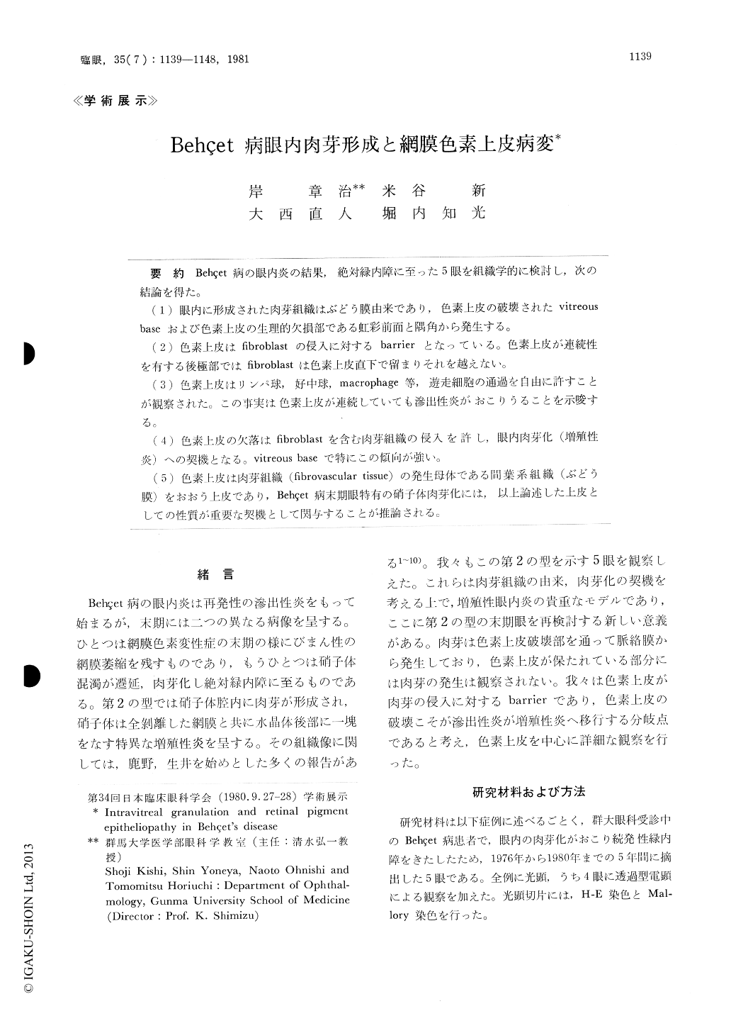
Behçet病の眼内炎の結果,絶対緑内障に至った5眼を組織学的に検討し,次の結論を得た。
(1)眼内に形成された肉芽組織はぶどう膜由来であり,色素上皮の破壊されたvitreousbaseおよび色素上皮の生理的欠損部である虹彩前面と隅角から発生する。
(2)色素上皮はfibroblastの侵入に対するbarrierとなっている。色素上皮が連続性を有する後極部ではfibroblastは色素上皮直下で留まりそれを越えない。
(3)色素上皮はリンパ球,好中球,macrophage等,遊走細胞の通過を自由に許すことが観察された。この事実は色素上皮が連続していても滲出性炎がおこりうることを示唆する。
(4)色素上皮の欠落はfibroblastを含む肉芽組織の侵入を許し,眼内肉芽化(増殖性炎)への契機となる。vitreous baseで特にこの傾向が強い。
(5)色素上皮は肉芽組織(fibrovascular tissue)の発生母体である間葉系組織(ぶどう膜)をおおう上皮であり,Behçet病末期眼特有の硝手体肉芽化には,以上論述した上皮としての性質が重要な契機として関与することが推論される。
We examined 5 eyes in the terminal stage of Behçet's disease by histological and electron micro-scopic means. Granulation tissue filled the vitre-ous cavity completely in each of the five eyes. We paid particular attention to the elucidation of the origin and the progression of granulation formation.
The granule tissue originated from the uvea through denuded retinal pigment epithelium (RPE) at the vitreous base, the anterior iris sur-face and the chamber angle. When the RPE still retained its continuity, fibroblasts could not invade the vitreous cavity from the choroid.
Copyright © 1981, Igaku-Shoin Ltd. All rights reserved.