Japanese
English
- 有料閲覧
- Abstract 文献概要
- 1ページ目 Look Inside
I.まえがき
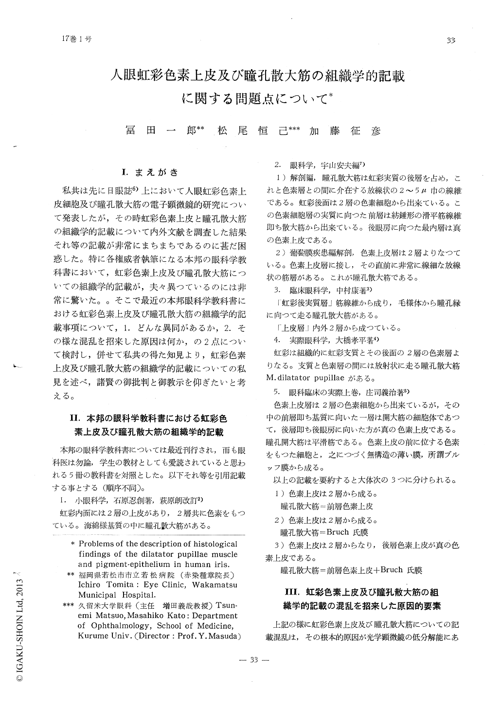
私共は先に日眼誌6)上において人眼虹彩色素上皮細胞及び瞳孔散大筋の電子顕微鏡的研究について発表したが,その時虹彩色素上皮と瞳孔散大筋の組織学的記載について内外文献を調査した結果それ等の記載が非常にまちまちであるのに甚だ困惑した。特に各権威者執筆になる本邦の眼科学教科書において,虹彩色素上皮及び瞳孔散大筋についての組織学的記載が,夫々異つているのには非常に驚いた。。そこで最近の本邦眼科学教科書における虹彩色素上皮及び瞳孔散大筋の組織学的記載事項について,1.どんな異同があるか,2.その様な混乱を招来した原因は何か,の2点について検討し,併せて私共の得た知見より,虹彩色素上皮及び瞳孔散大筋の組織学的記載についての私見を述べ,諸賢の御批判と御教示を仰ぎたいと考える。
As for the description histological findings of the dilatator pupillae muscle and pigment-epithelium of human iris, there are different names with different authors in our text-boo-ks. For classification of this matter, electron microscopic study performed in the human iris, and results are as follows,
1) The nucleus layer of the dilator pupillae muscle is the anterior pigment-epithelium of human iris, Therefore, it might be wise to call the posterior pigment-epithelium only the pigment-epithelium.
2) The Bruch's membrane consists of slender processes which rise from the nucleus layer of the dilator pupillae muscle.

Copyright © 1963, Igaku-Shoin Ltd. All rights reserved.


