Japanese
English
- 有料閲覧
- Abstract 文献概要
- 1ページ目 Look Inside
I.緒言
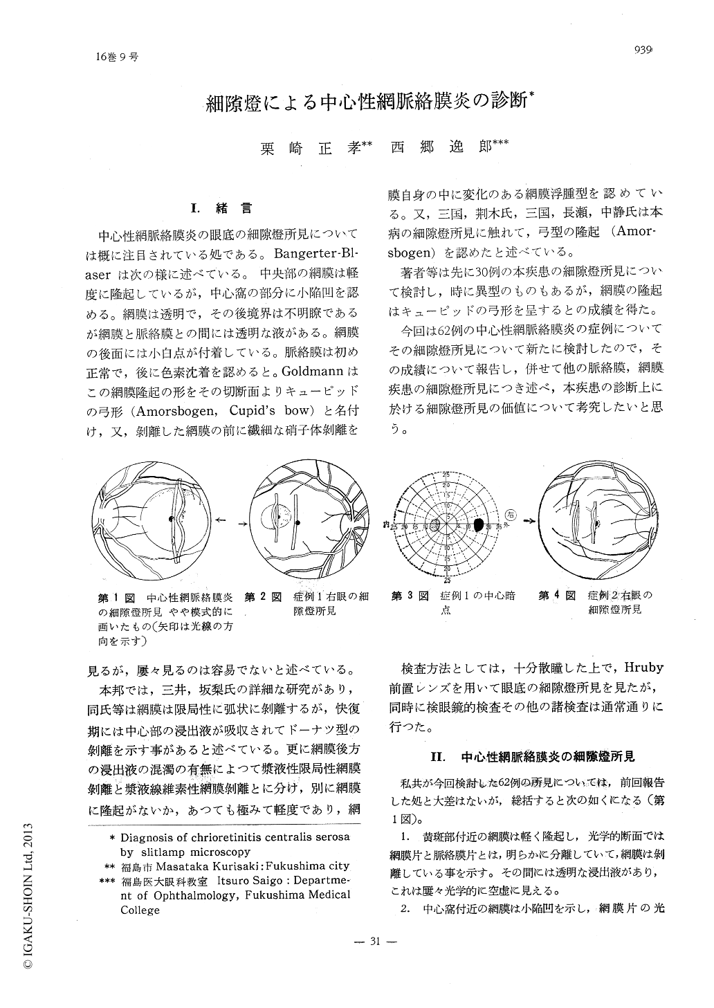
中心性網脈絡膜炎の眼底の細隙燈所見については概に注目されている処である。Bangerter-Bl—aserは次の様に述べている。中央部の網膜は軽度に隆起しているが,中心窩の部分に小陥凹を認める。網膜は透明で,その後境界は不明瞭であるが網膜と脈絡膜との間には透明な液がある。網膜の後面には小白点が付着している。脈絡膜は初め正常で,後に色素沈着を認めると。Goldmannはこの網膜隆起の形をその切断面よりキューピッドの弓形(Amorsbogen,Cupid's bow)と名付け,又,剥離した網膜の前に繊細な硝子体剥離を見るが,屡々見るのは容易でないと述べている。
本邦では,三井,坂梨氏の詳細な研究があり,同氏等は網膜は限局性に弧状に剥離するが,快復期には中心部の浸出液が吸収されてドーナツ型の剥離を示す事があると述べている。更に網膜後方の浸出液の混濁の有無によつて漿液性限局性網膜剥離と漿液線維素性網膜剥離とに分け,別に網膜に隆起がないか,あつても極みて軽度であり,網膜自身の中に変化のある網膜浮腫型を認めている。又,三国,荊木氏,三国,長瀬,中静氏は本病の細隙燈所見に触れて,弓型の隆起(Amor—sbogen)を認めたと述べている。
The authors have observed biomicroscopic findings of 62 cases of chorioretinitis centralis serosa. The results are as follows:
1. The retina of macula region is elevated slightly with a navel-like depression, corre-sponding to the fovea centralis, so that the optical section of the retina is shaped like a Cupid's bow (Goldman). We observed Cupid's bow shaped elevation of the retina, in 54 out of 62 cases.
2. Though in 16 out of 62 cases the retina was seen slightly opaque, the retina was normally transparent in the other cases.

Copyright © 1962, Igaku-Shoin Ltd. All rights reserved.


